[Update: November 18, 2014 Study published today – cause of Sea Star Wasting Syndrome a densovirus that has been present for at least 72 years? Why has it led to mass mortality now? What makes sense is that, like any virus, the incidents of “pathogenicity” depends on stressors (e.g. temperature change) and proximity of individuals. The virus has also been found in other echinoderms like urchins and sand dollars and it persists in sediment = can be transmitted by those vectors and there is the potential that the other echinoderms are/will be affected. See the study by Cornell University at the link below (lead author Ian Hewson). Includes “If SSaDV is the cause of the current SSWD event, it is unclear why the virus did not elicit wide disease outbreaks in the past during periods in which it was detected; however, there are several possible reasons why the current SSWD event is broader and more intense than previous occurrences. SSaDV may have been present at lower prevalence for decades and only became an epidemic recently due to unmeasured environmental factors not present in previous years that affect animal susceptibility or enhance transmission.” http://www.pnas.org/content/early/2014/11/12/1416625111.abstract]
I am very sad to report that Sea Star Wasting Syndrome is now on NE Vancouver Island.
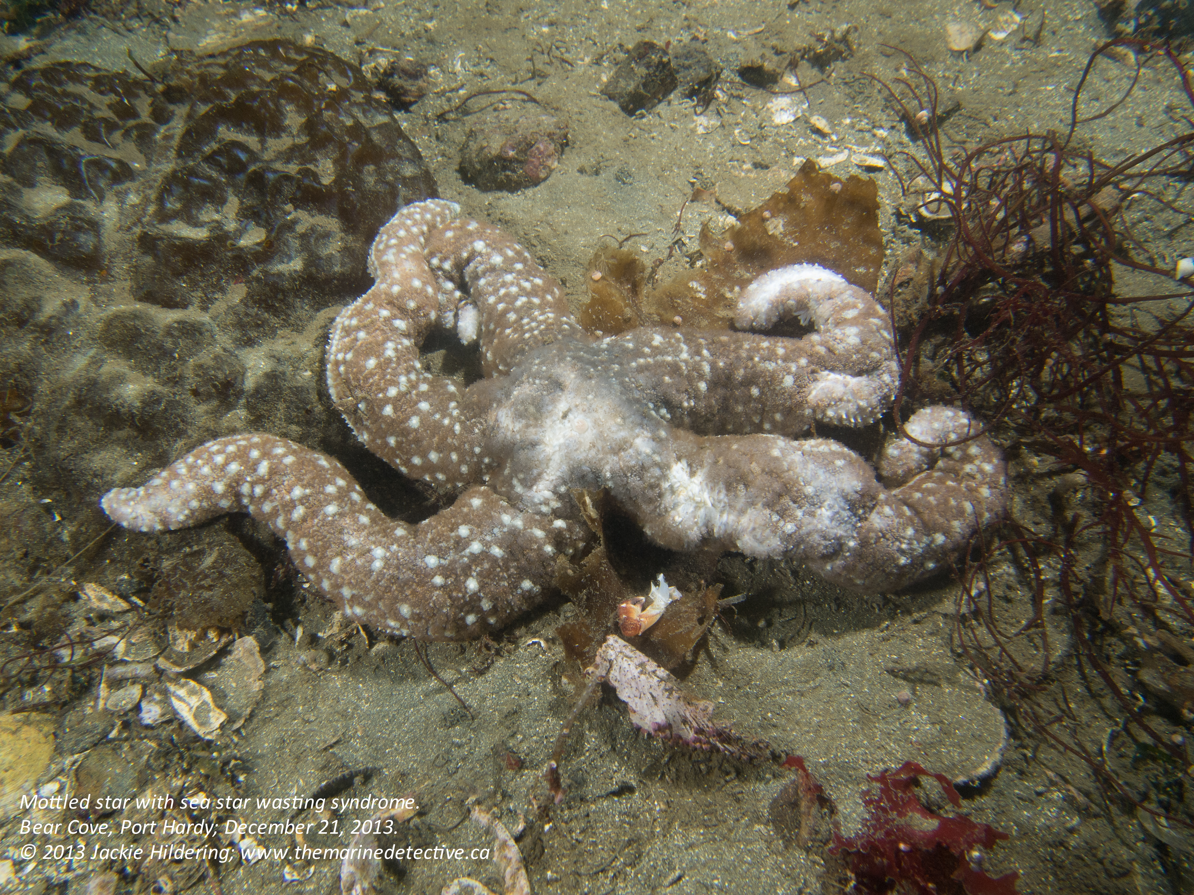
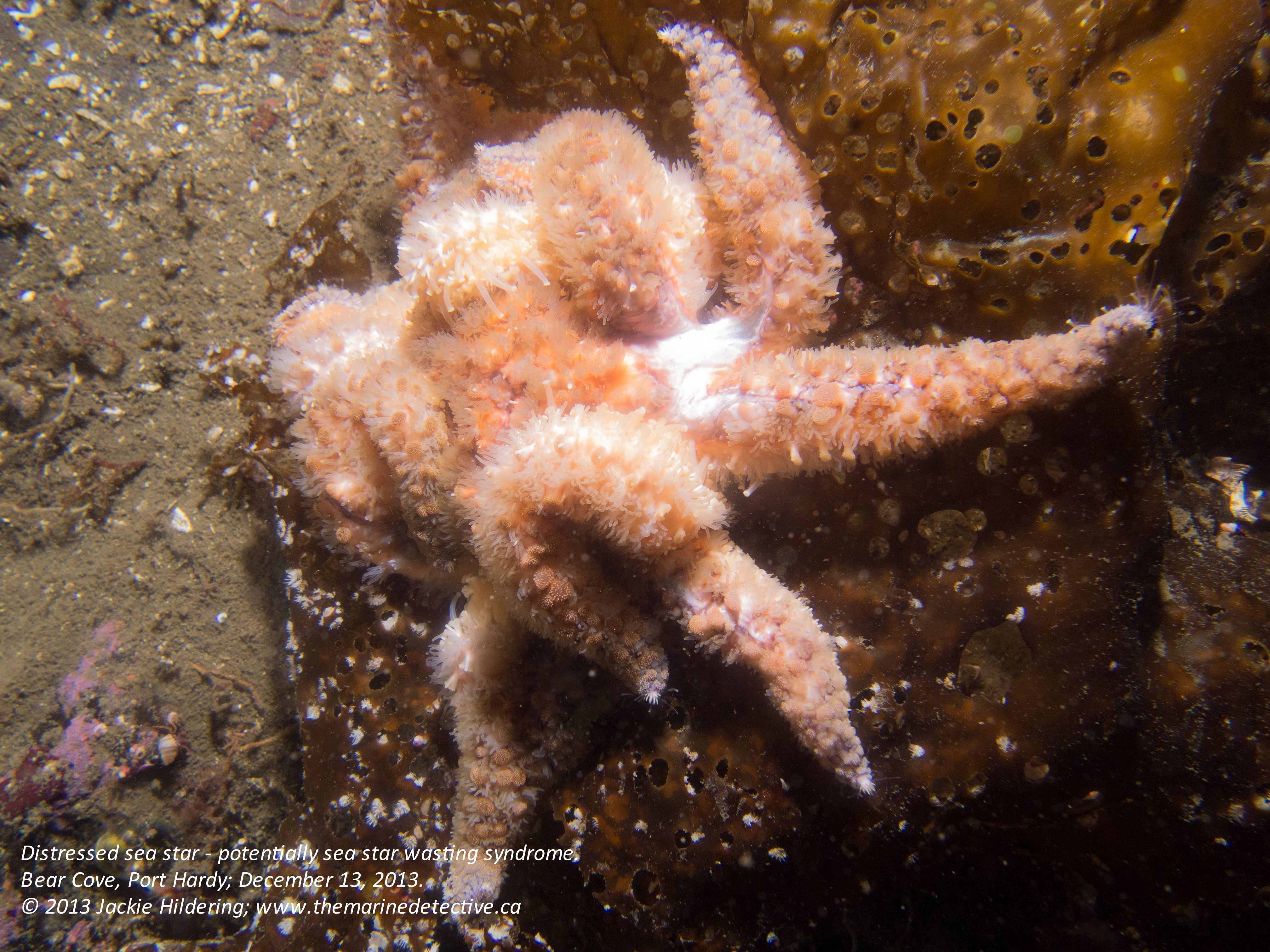
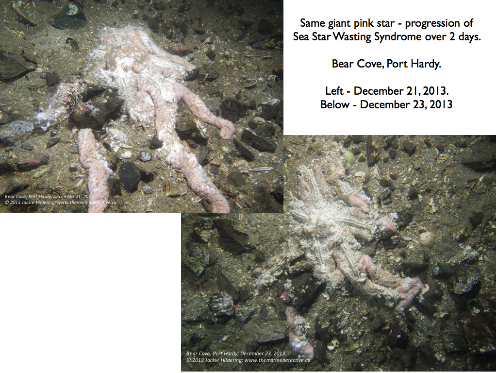
I first detected symptoms of the Syndrome at Bear Cove in Port Hardy on December 13th. Please see table at the end of this blog for how the species affected appears to be quite different from further to the south. Leather stars seem particularly affected and the Syndrome appears to advance much more slowly.
I have tried to think up a terrestrial analogy for what is happening to the sea stars so that non-divers might better get a sense of the weight and ecosystem importance of it. However, I can’t come up with a good terrestrial equivalent of an abundant group of highly visible, apex predators. My best attempt is to suggest you think of sea stars like birds of prey. Imagine what you would feel like if you were to notice they were dying, bodies deflating . . . then melting away and that this would progress very quickly and spread like wildfire.
The meltdown of sea stars was first detected in June 2013 in Washington State in ochre stars and in sunflower stars in Howe Sound (BC) in late August 2013 but has now been reported at sites from Alaska to the Mexican border.
The number of sea stars impacted is orders of magnitude greater than any previous known outbreak.
Most likely due to a pathogen (virus and or/bacteria). Cornell University is doing the genomic work. Toxins and environmental conditions have not been ruled out as the cause (or compounding factors).
If it is a pathogen, how quickly it spreads is influenced by the number of animals and if they are stressed. There are likely to be layers of stressors.
It has put forward by the scientific community that this could be a normal mechanism for overpopulation in sea stars.
While radioactivity certainly is an environmental stressor, the Fukushima Disaster has not been implicated in Sea Star Wasting Syndrome – really! From a January 30, 2014 Earth Fix article “scientists see Fukushima as an unlikely culprit because the die-offs are patchy, popping up in certain places like Seattle and Santa Barbara and not in others, such as coastal Oregon, where wasting has only been reported at one location.” (Also see January 19th, 2014 article “Half-Lives and Half-Truths – Discovering the truth about five of the most widespread myths of the Fukushima disaster” and scroll to the sources at the end of this article for scientific papers on the potential impacts of Fukushima).
The 1-minute time-lapse video below shows the progression of the Syndrome in a sunflower star over 7 hours.
Yep, it’s terrible.
However, I believe very strongly that, in attempting to raise awareness about marine environmental issues, I must always reflect on “what you can do”. If I do not, I contribute to the spread of a devastating human syndrome: Eco-paralysis. Symptoms include people becoming despondent, overwhelmed, and underactive in undertaking positive socio-environmental change, and often saying “It’s all hopeless”. The cause? This I do know. Eco-paralysis is the result of not seeing the common solutions between environmental problems.
Sea Star Wasting Syndrome is a solid indicator of how little we know about our life-sustaining oceans. It emphasizes the importance of humility and precaution in decision-making around the environment and how we are all empowered to reduce environmental stressors (with emphasis on reducing fossil fuel consumption and chemical use).
Having witnessed what I have over the last many weeks, I am all the more driven to assist others in (1) falling deeper in love with the NE Pacific Ocean by revealing the beauty below her surface and (2) feeling the joy that comes from creating change that is better for the environment and, therefore, ourselves.
I published this blog near the beginning of the onset of Sea Star Wasting Disease (SSWD) in 2013. It has been updated since 2013 with research developments. See the original blog at the end, which includes photos of the progression of SSWD.
Background: Since 2013, more than twenty species of sea star have been impacted by Sea Star Wasting Disease from Mexico to Alaska. There is local variation in the intensity of the disease and which species are impacted. It is one of the largest wildlife die-off events in recorded history. Sea stars contort, have lesions, shed arms and become piles of decay. Sunflower Stars (the world’s biggest sea star species) remain devastated with far-reaching impacts on kelp forests and the marine ecosystem.
Where to relay sea star data(of great value in understanding the survival, species impacted, range, and potentially, contributing factors of Sea Star Wasting Disease (SSWD):
August 4, 2025 – Very big breakthrough: After more than 10 years, the causative agent for Sea Star Wasting Disease (SSWD) has been found. Bacteria – Vibrio pectenicida (in the same family as bacteria that causes Cholera in humans).
Media release includes: “Now that scientists have identified the pathogen that causes SSWD, they can look into the drivers of disease and resilience. One avenue in particular is the link between SSWD and rising ocean temperatures, since the disease and other species of Vibrio are known to proliferate in warm water . . .”
Research paper includes: “Vibrio spp. have been coined ‘the microbial barometer of climate change’, because of the increasing prevalence of pathogenic species associated with warming water temperatures. Given that existing evidence indicates a relationship between increasing seawater temperature and SSWD incidence, an important next phase of research will be to empirically define this relationship, a goal now possible as a result of the identification of a causative agent.”
Prentice, M. B., Crandall, G. A., Chan, A. M., Davis, K. M., Hershberger, P. K., Finke, J. F., Hodin, J., McCracken, A., Kellogg, C. T. E., Clemente-Carvalho, R. B. G., Prentice, C., Zhong, K. X., Harvell, C. D., Suttle, C. A., & Gehman, A. M. (n.d.). Vibrio pectenicida strain FHCF-3 is a causative agent of sea star wasting disease. Nature Ecology & Evolution. ____________________________
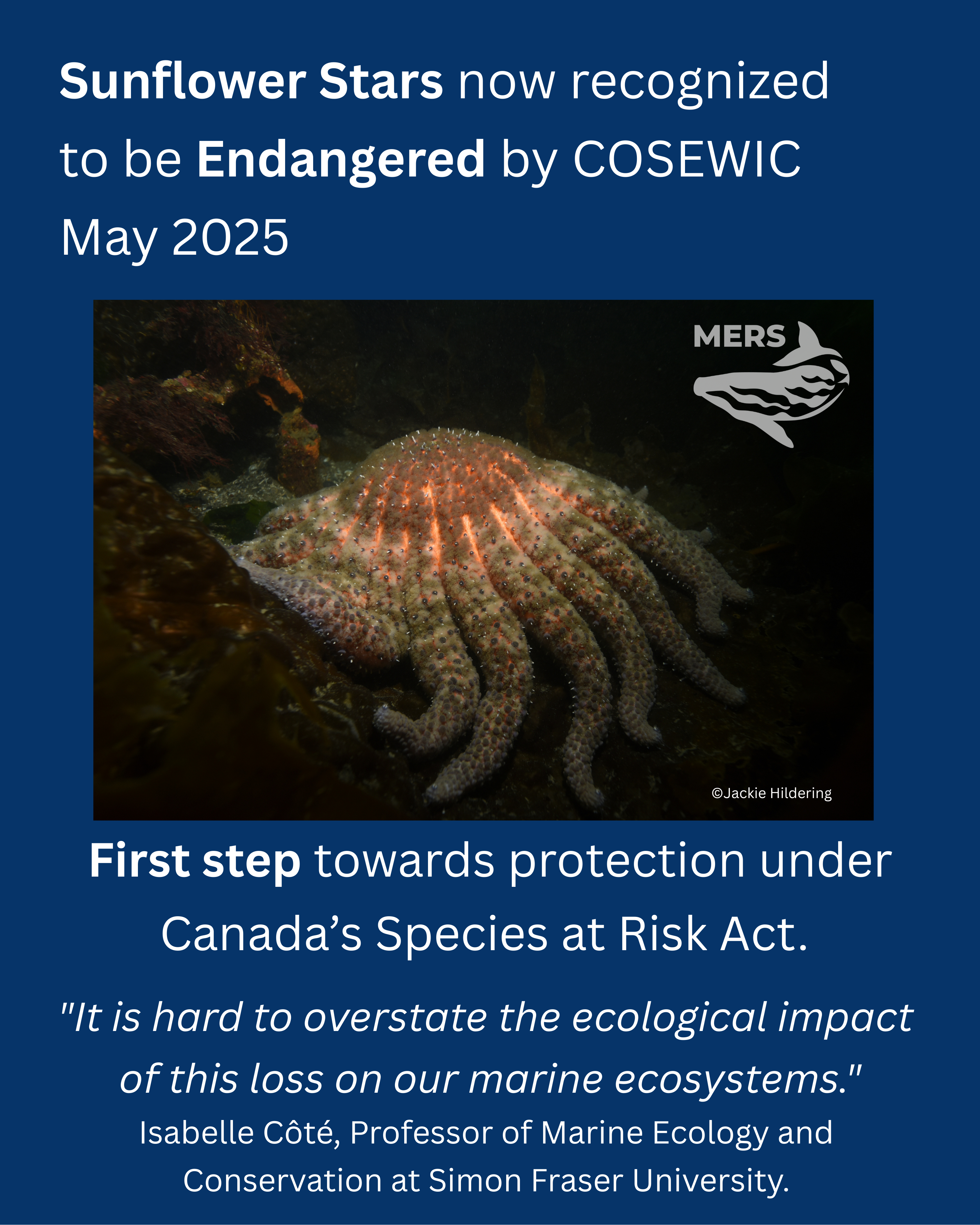
May 15, 2025 – Very important development: The Committee on the Status of Endangered Wildlife in Canada (COSEWIC) is recommending to the Government of Canada that Sunflower Stars be protected as an endangered species under Canada’s Species at Risk Act. This was decided at their May 8, 2025 meeting.
Why share the information about Sea Star Wasting Disease and put the effort into tracking and educating about the research?
It is often marine species that testify to environmental problems first, serving as indicators for the resources upon which we too depend. The hypothesis remains that the sea stars have succumbed in an unprecedented way because of changed ocean conditions (stressors). Too few of us realize the importance of sea stars in the ocean food web (see video below) let alone the importance of what they might be indicated about environmental health.
Quote from Drew Harvell, Cornell University professor of ecology and evolutionary biology who studies marine diseases: “these kinds of events are sentinels of change. When you get an event like this, I think everybody will say it’s an extreme event and it’s pretty important to figure out what’s going on . . . Not knowing is scary . . . If a similar thing were happening to humans, the Centers for Disease Control and Prevention would commit an army of doctors and scientists to unraveling the mystery.“
Below, January 30, 2019 video by the Hakai Institute re. Sunflower Stars and Sea Star Wasting Disease.
Research on Sea Star Wasting Syndrome in reverse chronological order:
Sunflower Stars are already recognized as Critically Endangered by the International Union for Conservation of Nature but this does not offer them protection in Canada or the US. In Canada, an “unsolicited assessment” has been provided to the Committee on the Status on Endangered Wildlife in Canada (COSEWIC) in hopes of expediting the protection of Sunflower Stars under Canada’s Species at Risk Act.
The March 15 announcement by NOAA includes: “While Sea Star Wasting Syndrome is not well understood, it appears to be exacerbated by rapid changes in water temperature, warmer ocean temperatures, and other physical stressors. Outbreaks are likely to recur as the climate continues to warm. Outbreaks may also be more frequent or spread more quickly . . . Populations of the species appear relatively more viable are in cooler, and possibly deeper, waters to the north, including Alaska, British Columbia, and the Salish Sea in the Pacific Northwest. Losses due to the syndrome in these waters were not as high as in more southerly waters.”
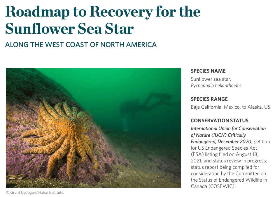
December 2022: Roadmap to recovery for the sunflower sea star (Pycnopodia helianthoides) along the west coast of North America. The Nature Conservancy (Heady et al). From the Executive Summary: “A sea star wasting disease (SSWD) event beginning in 2013 reduced the global population of sunflower sea stars by an estimated ninety-four percent, triggering the International Union for the Conservation of Nature (IUCN) to classify the species as Critically Endangered. Declines of ninety-nine to one hundred percent were estimated in the outer coast waters of Baja California, California, Oregon, and Washington. From the Salish Sea to the Gulf of Alaska, declines were greater than eighty-seven percent; however, there is uncertainty in estimates from Alaska due to limited sampling. A range-wide species distribution analysis showed that the importance of temperature in predicting sunflower sea star distribution rose over fourfold following the SSWD outbreak, suggesting latitudinal variation in outbreak severity may stem from an interaction between disease severity and warm waters. Given the widespread, rapid, and severe declines of sunflower sea stars, the continued mortality from persistent SSWD, and the potential for the disease to intensify in a warming future ocean, there is a need for a Roadmap to Recovery to guide scientists and conservationists as they aid the recovery of this Critically Endangered species . . . The area of greatest concern and need for immediate action common to all geographic regions is understanding disease prevalence and disease risk. Here we use the term “disease” to describe SSWD, also known as Sea Star Wasting Syndrome or Asteroid Idiopathic Wasting Syndrome, which affects some twenty species of sea stars and the cause(s) of which remain unknown and under debate in the literature. Much work is needed to improve our understanding of SSWD, the cause(s) of SSWD, how SSWD impacts wild sunflower sea stars, SSWD dynamics in a multi-host system, and to discover and develop measures to mitigate SSWD impacts and risks associated with conservation actions.”
December 29, 2021 – assessment report for the International Union for the Conservation of Nature = Gravem, S.A., W.N. Heady, V.R. Saccomanno, K.F. Alvstad, A.L.M. Gehman, T.N. Frierson and S.L. Hamilton. 2021. Pycnopodia helianthoides. IUCN Red List of Threatened Species 2021.
This research suggests that the pathogen is not a virus but a bacteria. The research puts forward that warmer oceans and increased organic matter appear to lead to increases in specific bacteria (copiotrophs) that then use up the oxygen at the interface of the sea star and the bacteria, and the sea stars can’t breathe. The hypothesis includes that “more heavily affected species were rougher and therefore had a much larger boundary layer (the layer at the animal-water interface) than those species which were less affected.”
Quote from lead author: “The main takeaway is the speed with which a multi-host infectious disease can cause decline in the most susceptible host [Sunflower Stars] and that warming temperatures can field bigger impacts of disease outbreaks.” Abstract includes: “Since 2013, a sea star wasting disease has affected >20 sea star species from Mexico to Alaska. The common, predatory sunflower star (Pycnopodia helianthoides), shown to be highly susceptible to sea star wasting disease, has been extirpated across most of its range. Diver surveys conducted in shallow nearshore waters (n = 10,956; 2006–2017) from California to Alaska and deep offshore (55 to 1280 m) trawl surveys from California to Washington (n = 8968; 2004–2016) reveal 80 to 100% declines across a ~3000-km range. Furthermore, timing of peak declines in nearshore waters coincided with anomalously warm sea surface temperatures. The rapid, widespread decline of this pivotal subtidal predator threatens its persistence and may have large ecosystem-level consequences.”
The paper’s discussion includes: “Cascading effects of the P. helianthoides loss are expected across its range and will likely change the shallow water seascape in some locations and threaten biodiversity through the indirect loss of kelp. P. helianthoides was the highest biomass subtidal asteroid across most of its range before the Northeast Pacific SSWD event. Loss or absence of this major predator has already been associated with elevated densities of green (Strongylocentrotus droebachiensis), red (Mesocentrotus franciscanus), and purple urchins (Strongylocentrotuspurpuratus) across their range, even in regions with multiple urchin predators. Associated kelp reductions have been reported following the outbreak . . . SSWD, the anomalously warm water, P. helianthoides declines, and subsequent urchin explosions . . . have been described as the “perfect storm.” This “storm” could result not only in trophic cascades and reduced kelp beds but also in abalone and urchin starvation.”
Sunflower Stars are of great ecological importance in maintaining kelp forests. Burt et al in 2018 quantifies the importance of Sunflower Stars in maintaining kelp forests. Sunflower Stars feed on Green Urchins which graze on kelp. Findings included that the decline of Sunflower Stars “corresponded to a 311% increase in medium urchins and a 30% decline in kelp densities”. The loss of kelp forests can impact many other ecologically and commercially important species that relay upon them as habitat and food. Note too that our reliance on kelp forests includes oxygen production and carbon dioxide buffering.
This research, specifically on Ochre Stars, found that the genetic makeup of the species has changed since the outbreak. Young Ochre Sea Stars are more similar genetically to adults who survived than to those who succumbed. This “may influence the resilience of this keystone species to future outbreaks”. The findings of an additional March 2018 paper (Miner et al) include ” . . . we documented higher recruitment of P. ochraceus [Ochre Stars] in the north than in the south, and while some juveniles are surviving (as evidenced by transition of recruitment pulses to larger size classes), post-SSWD survivorship is lower than during pre-SSWD periods.
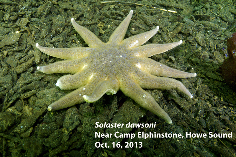
Sunflower Star with Sea Star Wasting Syndrome. Tissue wastes away. Legs often break off and crawl away briefly before rotting away. Photo – Neil McDaniel; http://www.seastarsofthepacificnorthwest.info
___________________________________________
The content below is from my original blog November 10, 2013:
There has already been much reporting on the gruesome epidemic spreading like wildfire through several species of sea star in the NE Pacific Ocean.
“Sea Star Wasting Syndrome” is incredibly virulent and is causing the mass mortality of some sea star species in British Columbia and beyond. “Sea stars go from “appearing normal” to becoming a pile of white bacteria and scattered skeletal bits is only a matter of a couple of weeks, possibly less than that” (Source #1).
What I have strived to do is bundle the state of knowledge, relying heavily on the expertise of two extraordinary divers and marine naturalists: (1) Neil McDaniel, marine zoologist and underwater photographer / videographer who maintains a website on local sea stars and has put together A Field Guide to Sea Stars of the Pacific Northwest, and (2) Andy Lamb, whose books include Marine Life of the Pacific Northwest.
I am hoping that kayakers, beach-walkers and fellow divers will help monitor and report on the spread of the disease but I am also hoping that all of us may learn from this tragedy that has impacted “one of the most iconic animals on the coast of British Columbia . . . more abundant and diverse in our waters than anywhere else in the world” (Source #3).
Sea Star Wasting Syndrome reminds us of the fragility of ocean ecosystems; how very quickly disease could spread in the ocean; and how we are all empowered to reduce stressors that increase the likelihood of pathogens manifesting as disease (e.g. climate change) or even that pathogens enter the environment (e.g. sewage).
Species impacted?
High mortalities (note that the first 4 are members of the same family – the Asteriidae):
Sunflower star (Pycnopodia helianthoides) hardest hit in southern British Columbia. From communication with Neil McDaniel ” . . .so far I estimate it has killed tens, possibly hundreds of thousands of Pycnopodia in British Columbia waters.”
Update January 21st, 2014: Possibly: Rose star (Crossaster papposus) – I have noted symptoms in this species on NE Vancouver Island as has Neil McDaniel in S. British Columbia).
Symptoms and progression of SSWD:
Neil McDaniel shared the following 7 images for the progression of the disease in Sunflower Stars [Source #2 and #14]. See the end of this blog item for images showing symptoms in other sea star species as well as a 1 minute time-lapse clip showing the progression of the syndrome in a Sunflower Star over 7 hours. [Note that the progression of the Syndrome on NE Vancouver Island appears that it may be different from what has been observed further to the south.]
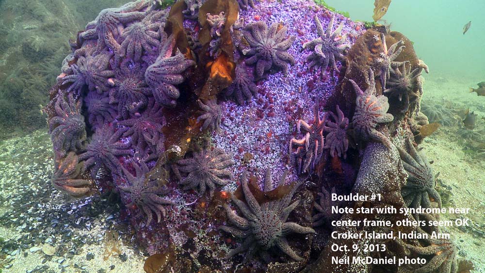
1. In this image most of the Sunflower Stars appear healthy “other than one just right of center frame is exhibiting the syndrome, looking “thinned-out” and emaciated.”
3. This image “shows how the body wall begins to rupture, allowing the gonads and pyloric caeca to spill out.”
As the animals become more stressed, they often drop several rays (which wander off on their own for a while). At this point the body wall becomes compromised and the pyloric caeca and/or gonads may protrude through lesions. As things progress, the animals lose the ability to crawl and may even tumble down steep slopes and end up in pile at the bottom. Soon after they die and begin to rot
5. As things progress, the animals lose the ability to crawl [and hold grip surfaces] and may even tumble down steep slopes and end up in pile at the bottom. Soon after they die and begin to rot.
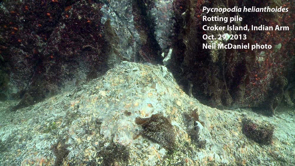
6. The bacteria Beggiatoa then takes over and consumes all of the organic matter, leaving a scattering of skeletal plates on the bottom. The syndrome develops quickly and in only one to two weeks animals can go from appearing healthy to a white mat of bacteria and skeletal plates
The 1-minute time-lapse video below shows the progression of the Syndrome in a sunflower star over 7 hours.
Cause(s)? To date, the cause(s) have not yet been identified. Scientific opinion appears to be that most likely the cause is one or more viruses or bacteria. As with any pathogen (like the flu virus), the expression of a pathogen as disease is influenced by the number and proximity of individuals and could be exacerbated by environmental stressors.
Has this happened before? Never to this large a scale. “Although similar sea star wasting events have occurred previously, a mortality event of this magnitude, with such broad geographic reach has never before been documented.” (Source #17).
“Southern California in 1983-1984 and again (on a lesser scale) in 1997-98” (Source #4 and #13)
Florida (Source #5).
Update November 30: Sunflower die offs [on much smaller scale] have been noted in the past in Barkley Sound. In 2008 ochre star die offs were documented in Barkley Sound. In 2009 Bates et. al. reported on this and observed that the prevalence of disease “was highly temperature sensitive and that populations in sheltered bays appeared to sustain chronic, low levels of infection.” (Source #14 and #15).
“Similar events have occurred elsewhere over the last 30 years. Sea stars have perished in alarming numbers in Mexico, California and other localities” (Source #2).
“In July, researchers at the University of Rhode Island reported that sea stars were dying in a similar way from New Jersey to Maine . . a graduate student collected starfish for a research project and then watched as they “appeared to melt” in her tank” (Source #5).
Shellfish Health Report from the Pacific Biological Station (DFO) conducted on 1 morning sun star and 7 sunflower stars collected on October 9, 2013 at Croker Island, Indian Arm; case number 8361.
Bates AE, Hilton BJ, Harley, CDG 2009. Effects of temperature, season and locality on wasting disease in the keystone predatory sea star Pisaster ochraceus. Diseases of Aquatic Organisms Vol. 86:245-251 http://www.ncbi.nlm.nih.gov/pubmed/20066959
Morning sun star with lesions indicating the onset of sea star wasting syndrome. Photo and descriptor – Neil McDaniel; http://www.seastarsofthepacificnorthwest.info Click to enlarge.
I am typing with salt still encrusted to my face and hair. I really should warm up from my dive and wash off the NE Pacific before sharing this with you but this is the kind of story you want to shout from the seamount tops. However, be warned, there is a bit of a dark side to the story too.
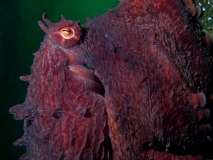
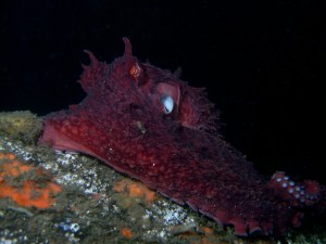
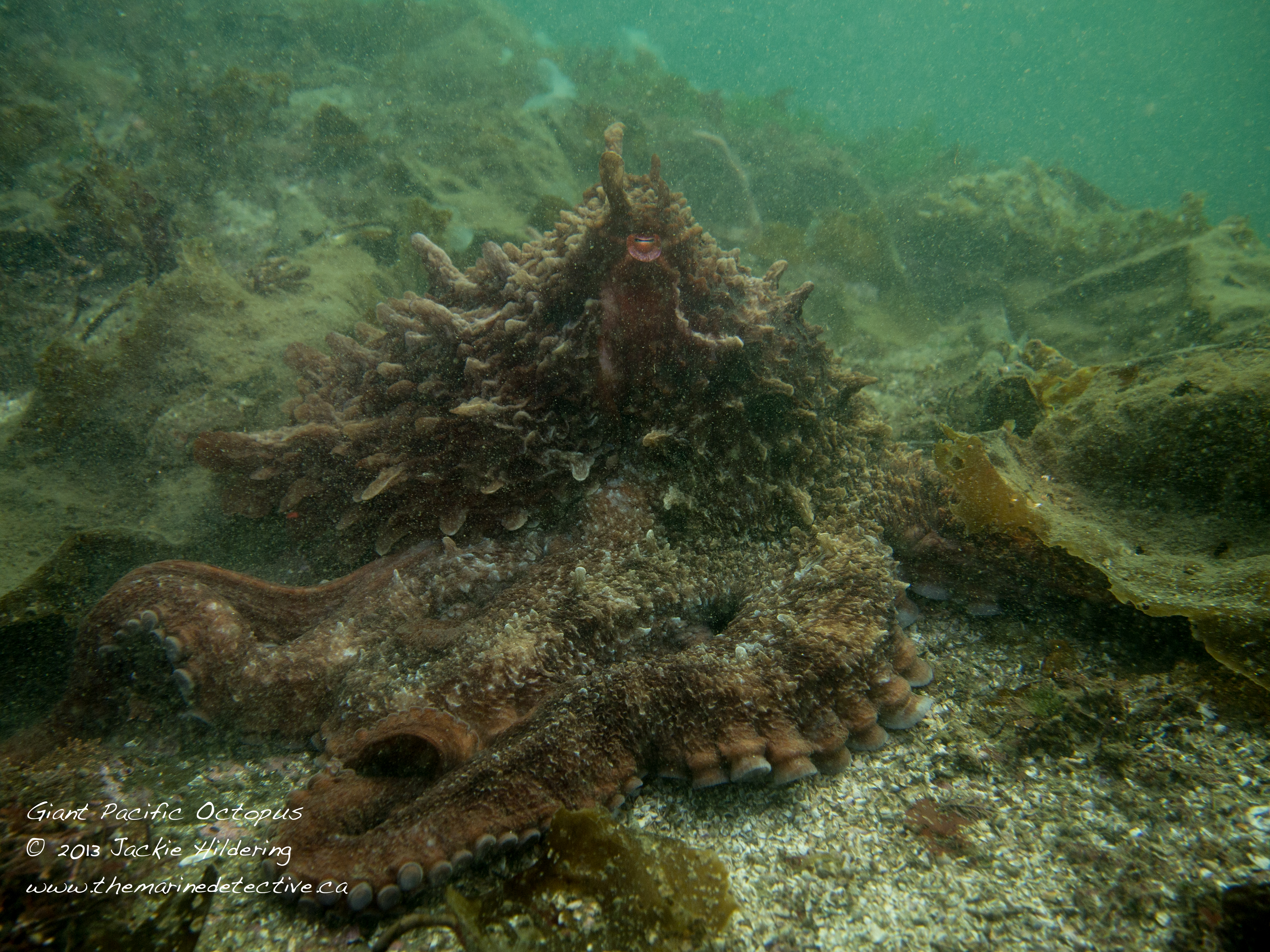
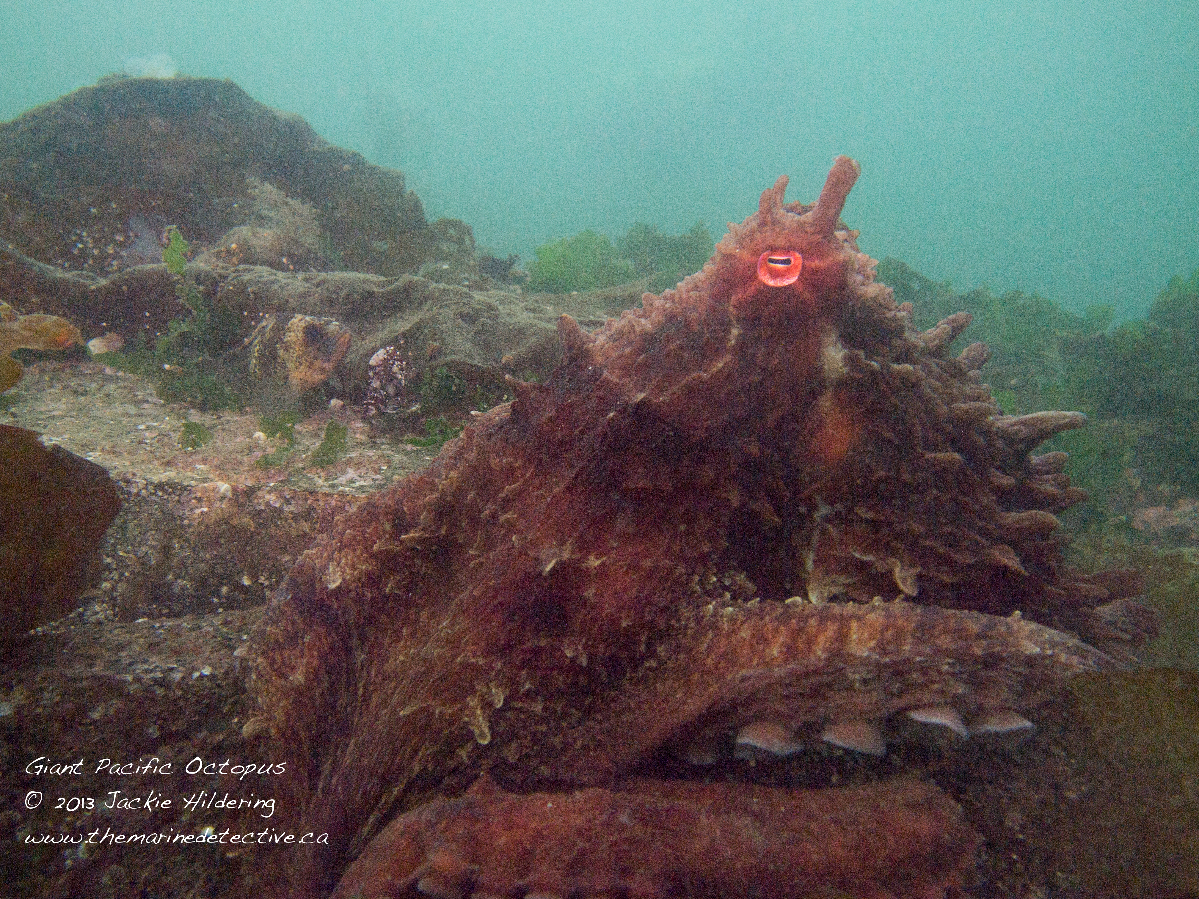
Today, while doing a shore dive in Port Hardy with the intention of surveying the health of sea stars*, I had the most wondrous experience I have ever had with not one, but two giant Pacific octopuses.
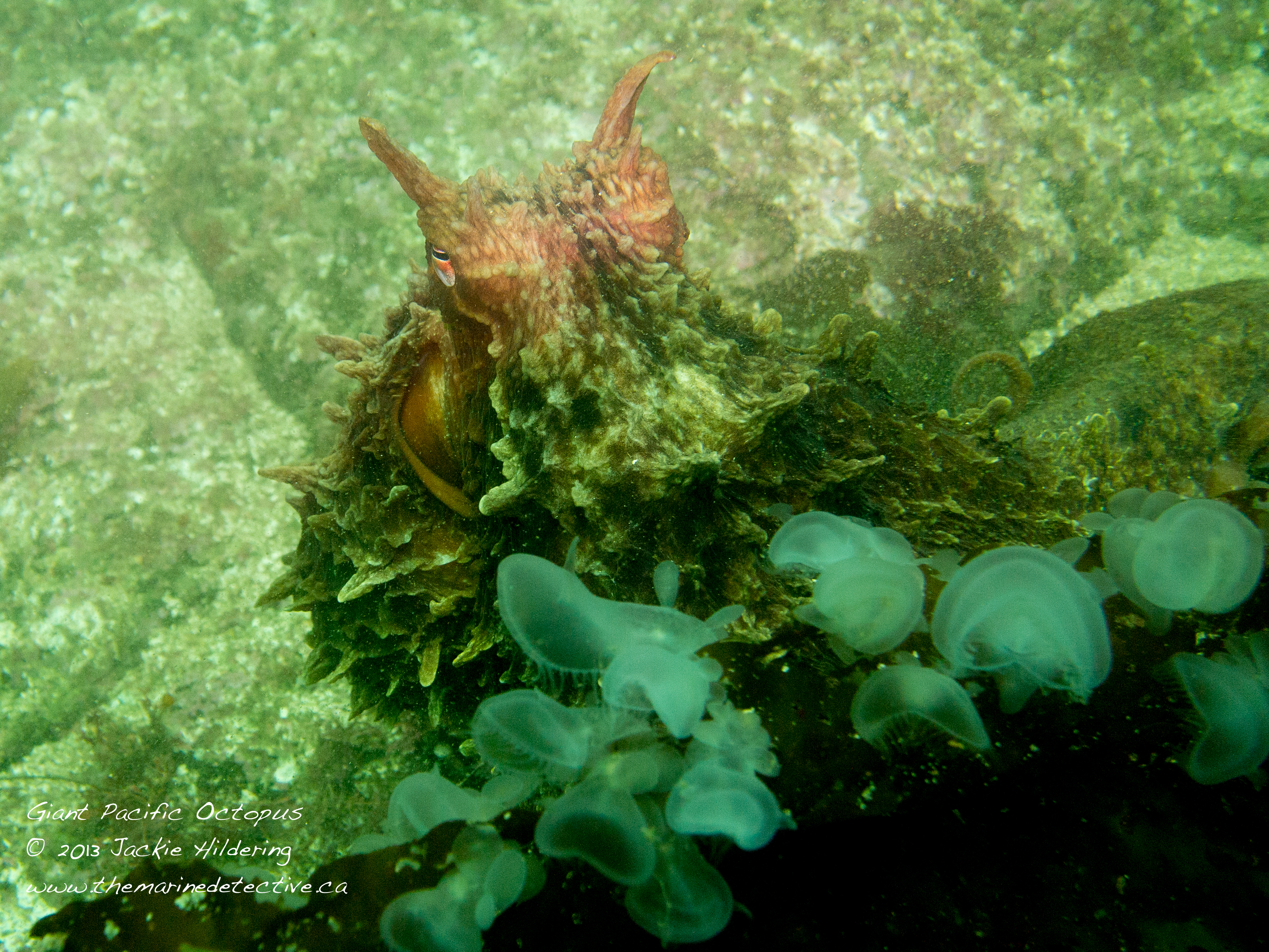
While photographing a sea star I must have disturbed the first octopus because when I looked down, wondering what had caused a massive disruption of hooded nudibranchs from the kelp, there she/he was in full glory – posturing to show me his/her impressive size, hooded nudibranchs undulating all around.
I even ended up with a hooded nudibranch stuck to my mask, which I gently shook off as I am a poor surrogate for kelp!
After I recovered from the shock of this all and mumbled an apology in the guilt of triggering the chaos, I looked at the octopus for a bit . . . and she/he looked at me. We both settled down, apparent in the case of the octopus in that he/she was no longer posturing and reverted to camouflage colours rather than alarm vibrance.
After some minutes, the assessment appeared to be made by this sentient being that I was not a risk; and that there was no need to hide (nor ink!). As a result, for half an hour I was able to (respectfully) follow along as the octopus hunted.
I was allowed to learn about hunting strategy and see how the colour and texture changed as it moved and how the mantle would flash white as it pounced upon prey.
The only thing that stopped this deeply awe-inspiring experience was that dive buddy, Alex Spicer, found a second octopus in the open!
This much smaller octopus was using giant kelp like a hammock.
The divers among you know what a rare gift it is to find one, let alone two, (unhabituated) octopuses out of their dens, certainly during daytime. The underwater photographers and videographers among you would be twitching all the more, knowing what an incredible opportunity this offers to capture the beauty of these giant wonders.
Here’s the dark side. Thankfully it is a literal dark side. My strobes (flashes) didn’t work properly and it was my own doing. It’s been a crazy week of work and, in the flurry resulting from wanting to fit in a dive, I forgot the cables that hook the strobes to the camera.
Yes, I was given what may be the opportunity of a lifetime but failed to fully capture the beauty of it, leaving you with only the grainy images below. However, I got to fully live the experience and had anything changed in the course of events that led to today’s dive, likely I wouldn’t have been octopused at all.
I hope the images are still enough to illuminate the joy and wonder I felt.
[Be sure you scroll down for the photo of the little guy in the kelp hammock!]
How does studying whale acoustics lead to increased knowledge about the depth range of nudibranchs?
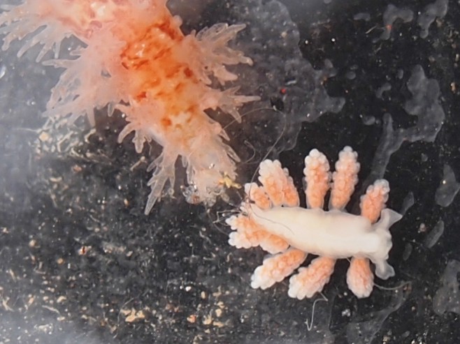
Just a little more is now known about the orange doto’s depth range. Photo: Hildering.
Let me take you deep and share an experience from my recent time offshore in the eastern North Pacific on a DFO cetacean survey.
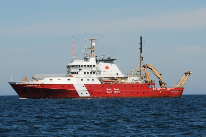
This is the Canadian Coast Guard Ship – the J.P. Tully.
CCGS J.P. Tully. Photo: Hildering
Among the offshore science expeditions undertaken upon the Tully, are surveys by DFO’s Cetacean Research Program. These line transect studies provide an estimate of cetacean abundance, as well as an opportunity to ID individual whales and collect feeding and genetic information. The knowledge about abundance and location is of particular importance for the large whales that were hunted so intensely and require protection under Canada’s Species at Risk Act.
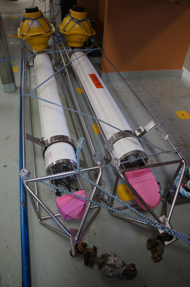
These are Autonomous Underwater Recorders for Acoustic Listening (AURAL-M2s).
AURAL-M2s. Photo: Sheila Thornton.
AURALs are hydrophones that can be deployed to 300 m, making time-spaced recordings (e.g. 15 minutes every hour) for up to a year. Such acoustic monitoring is a very important supplement to the cetacean vessel surveys. The AURALs are of course placed very strategically, in remote, offshore locations. By passively recording whale calls, the AURALs can provide information about the location and seasonality of whale species which may aid in determining critical habitat.
The AURALs are a wonder of technology. It is of course no problem to get something to the bottom of the ocean but, getting it back to the surface so you can retrieve your equipment and data is not so simple. It is achieved with an acoustic release (“D” in the diagram below). Once the vessel is positioned so that there is no chance of the device coming up under it, a sound signal is sent to the device and the AURAL releases from its anchor and floats to the surface thanks to the big yellow buoy.
AURAL-M2. Click to see an enlarged, labeled schematic on the Multi-Electronique webpage.
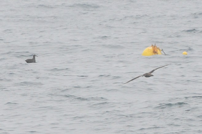
These are two perplexed black-footed albatrosses! A big yellow orb has just popped up to the surface as a result of the acoustic release signal. This AURAL was at 226 m depth at the Bowie Seamount, 180 km west of Haida Gwaii. It had been there for a year.
Black-footed albatross just after the buoy with the AURAL recording device came up from 226 m.
Here, the highly skilled Coast Guard crew get the AURAL back aboard the ship so that the data can be retrieved and, ultimately, analyzed for whale vocals.
Coast Guard deck crew expertly retrieves the AURAL. Photo: Hildering
But, there was also a year’s worth of growth on the buoy and who knows what you might find . . .
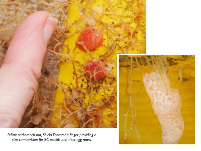
Nudibranchs! Three species found and even one species with eggs!
3 nudibranch species on the AURAL that had been at 226 m. BC aeolid; bushy-backed nudibranch and orange doto. Click to enlarge. Photo: Hildering.
Top: BC aeolid (Catriona columbiana to 1.5 cm); eggs also found.
By examining the AURAL that had been at 226 m, it confirms that these 3 species of nudibranch have a range to at least that depth.
Sheila Thornton (marine mammal researcher and fellow nudibranch nut) providing a size comparison for the BC aeolids and their egg masses that were found on the AURAL. Click to enlarge. Photo: Hildering
I shared the find with those who have nudibranch expertise much greater than my own (Dave Behrens via Andy Lamb) and learned that for two of the species, there had been no previous record for them at this depth.
It has long been known that some nudibranch species range to depths of at least 700 m. However, you can imagine what a a challenge it is to get species specific depth information. We camera carrying scuba divers can’t help beyond 40 m depth (deeper if diving with mixed gases).
So it’s not a big scientific discovery. Compared to the data the AURAL will reveal about endangered whales, it’s just a sea-slug-sized discovery.
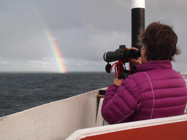
This is me – back on survey duty looking for much bigger organisms but delighting in how collecting data to help save whales, led to learning a bit more about the little guys.
Spotter duty on the DFO Cetacean Program’s offshore survey. July 2013. Christie McMillan photo.
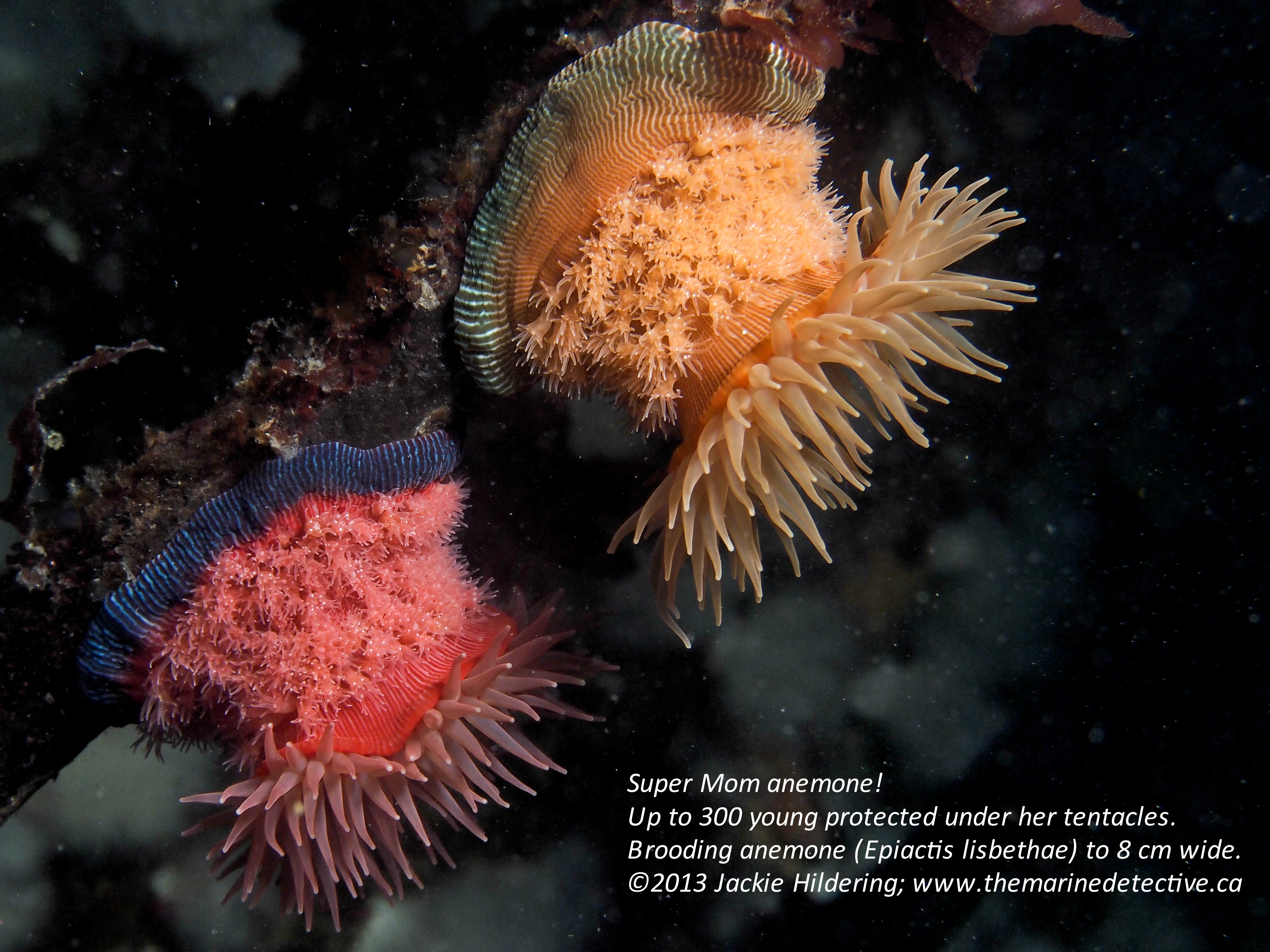
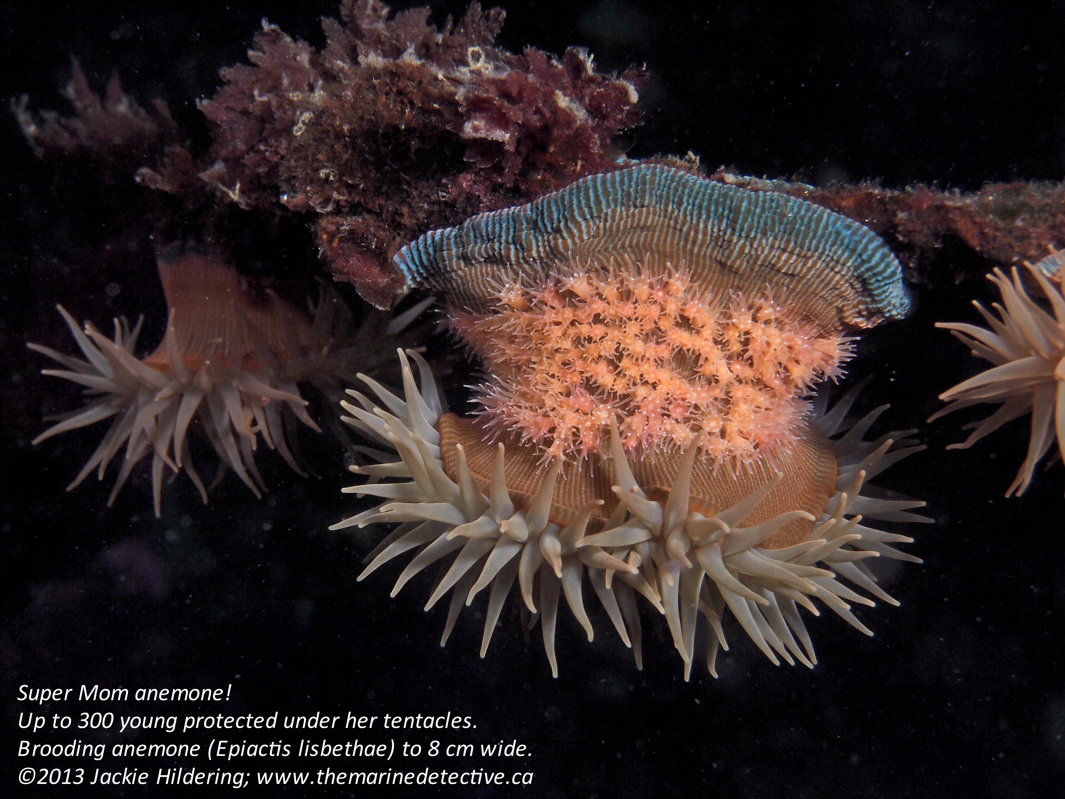
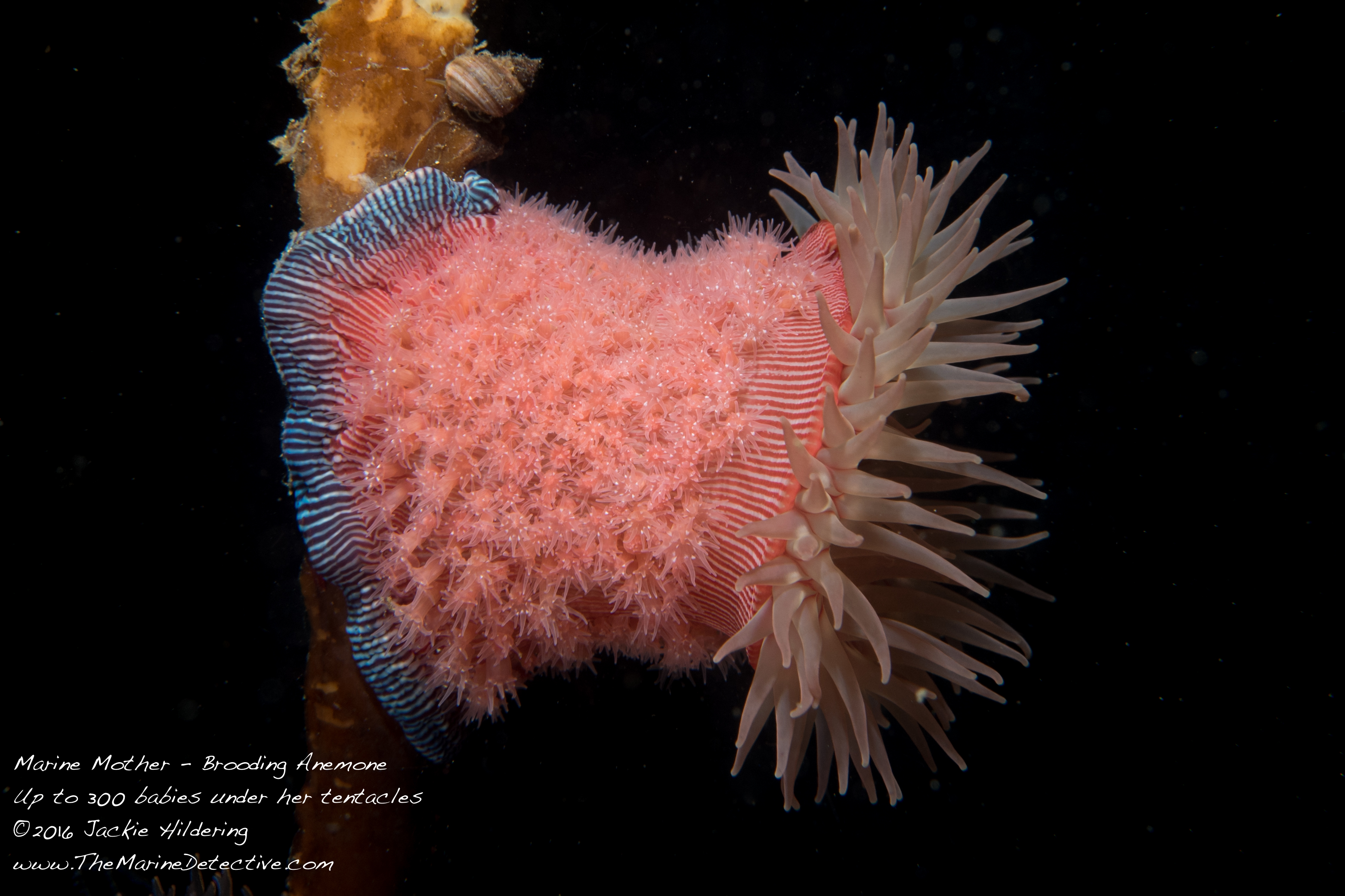
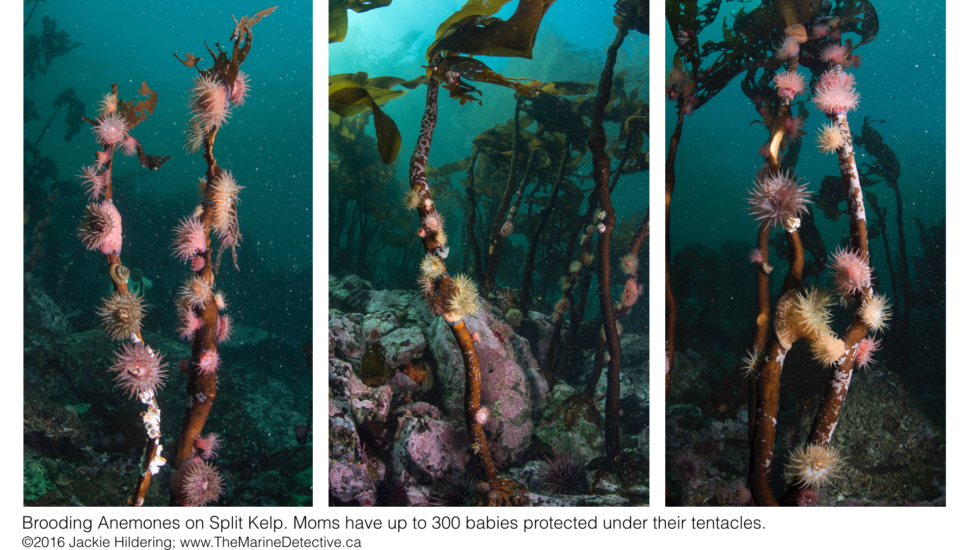
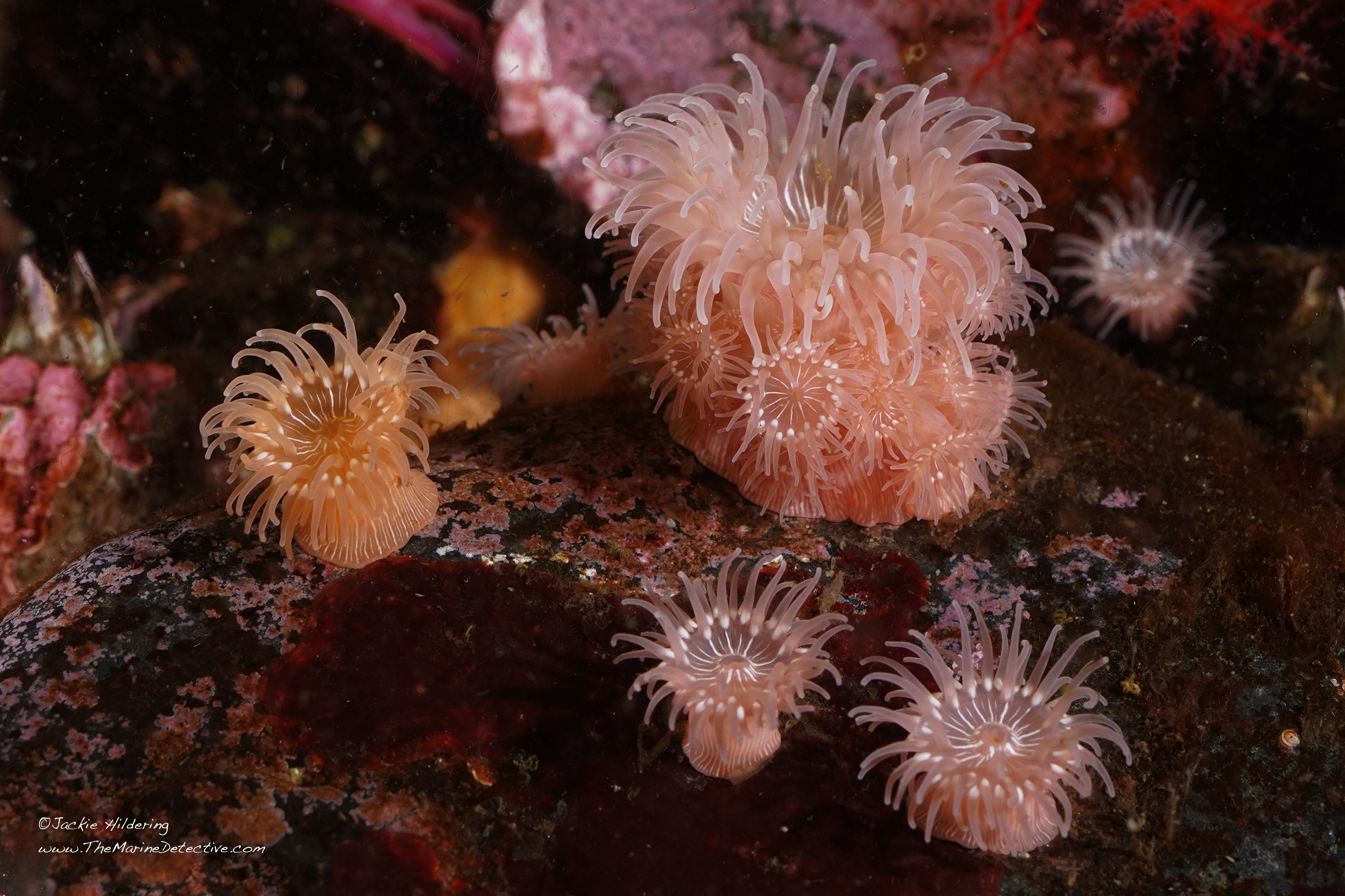
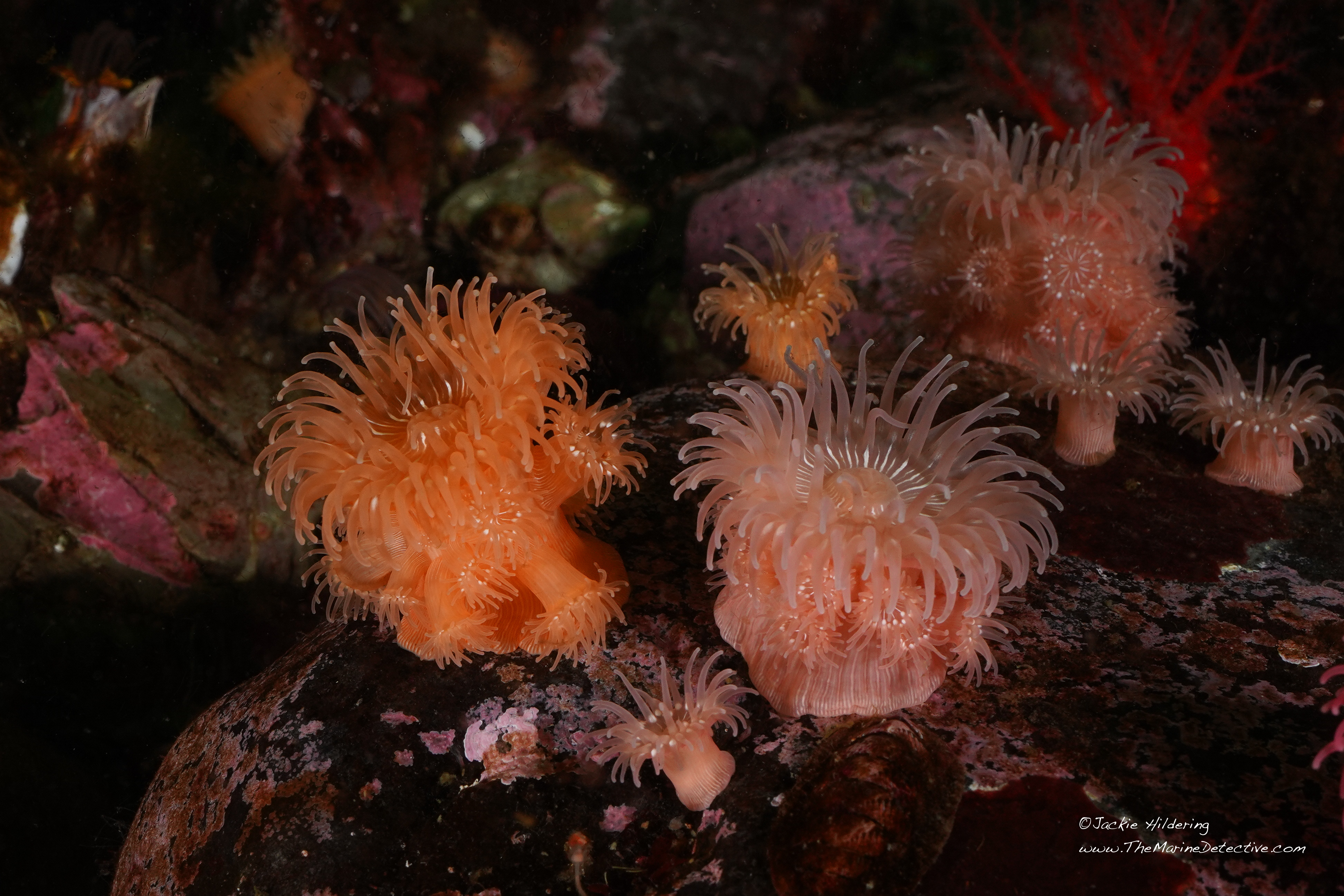
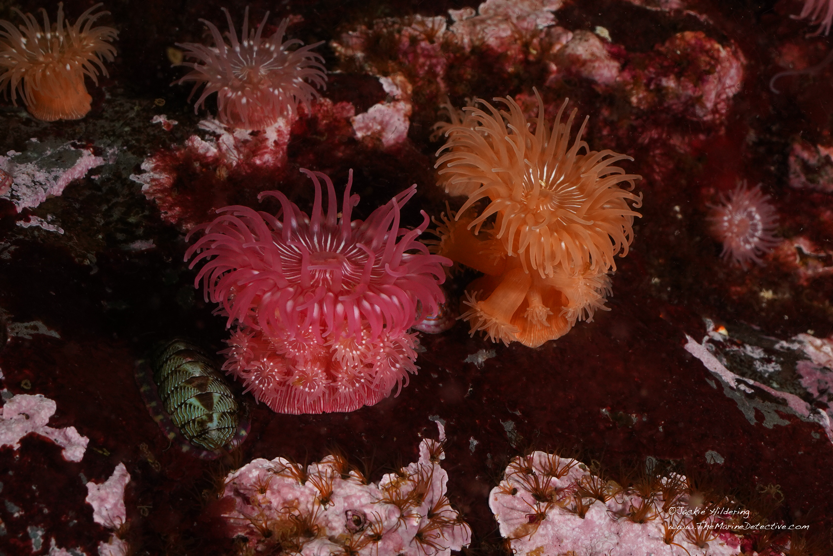
This is a Brooding Anemone (Epiactis lisbethae to 8 cm across).
She may not have a backbone but she’s a Super Mom!
As many as 300 young can be clustered around her in up to 5 rows, benefitting from the protective canopy of her tentacles which contain stinging cells (nematocysts). The offspring remain here until big enough to stand a good chance of surviving on their own. They then crawl toward independence, claiming their own piece of the ocean bottom.
I am awestruck by this species’ beauty and reproductive strategy. It is also a reminder of how little we know about marine species that the Brooding Anemone was not recognized as a distinct species until fairly recently (1986), and it still so often gets confused with the Proliferating Anemone (Epiactis prolifera).
I share my marine “detectiving” about this species with you to provide a further example of how extraordinary our marine neighbours are and maybe, thereby, help inspire greater conservation efforts.
But yes, the timing of the blog is no accident. It may be that reflection upon an anemone Super Mom stimulates thought about our human mothers – just in time for Mother’s Day.
So here goes . . . bear with me as I build to clarifying the reproduction of our featured species.
Anemones have many reproductive strategies.
For many species, reproduction can be asexual as well as sexual with strategies like budding off offspring; splitting into two; or pedal laceration where a torn piece of the bottom of the anemone can grow into another anemone!
Some species are hermaphrodites with highly diverse ways by which offspring develop into adults.
In species that have separate sexes, many are broadcast spawners where Mom and Dad release their eggs and sperm into the ocean around them. Fertilization and development thereby happens in the water column.
Then, for only some 20 species of the world’s more than 800 kinds of anemone, there are those in which the female captures the males’ sex cells as they drift by and draws them into her digestive cavity to fertilize her eggs. She “broods’ her young.
Some such anemone species are internal brooders. The young develop inside Mom until they hatch and are expelled into the water column as planktonic larvae.
But then there’s Super Mom – the Brooding Anemone (Epiactis lisbethae). She’s an external brooder.
After she has fertilized the eggs inside her digestive cavity with the sperm she has captured, the young develop inside her until they hatch into planktonic larvae. THEN, they swim out of her mouth, settle on her body under the tentacles and grow into little anemones that feed themselves.
When the offspring are big enough to stand a good chance of survival without the protection of Mom’s tentacles, they shuffle away to independence, leaving space for next season’s young.
The brooding anemone’s young are all of the same generation and are therefore all about the same size.
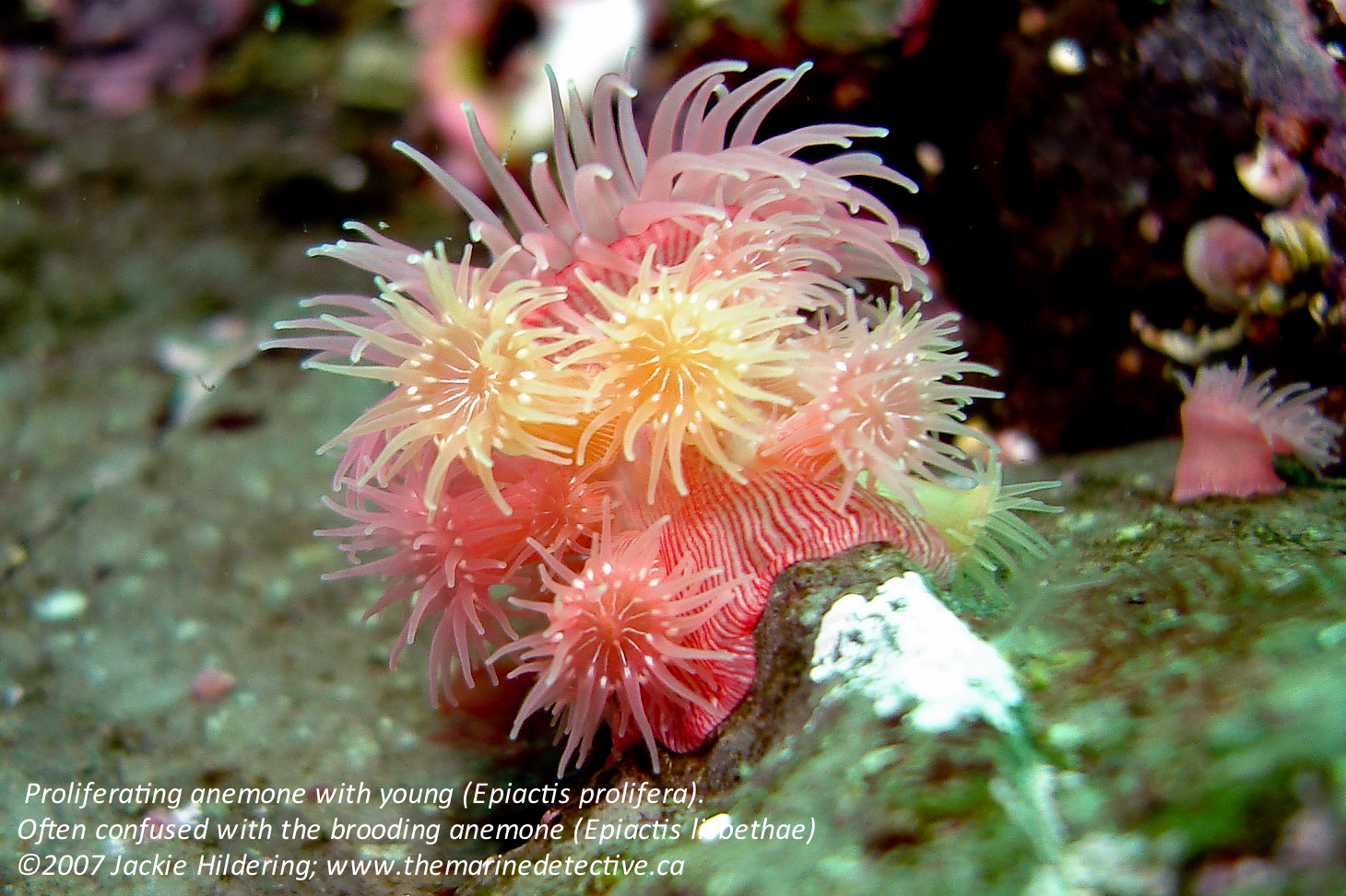
However, there is a second externally brooding anemone species in the eastern North Pacific where you most often see young of different sizes huddled under Mom’s tentacles. This species – the Proliferating Anemone (Epiactis prolifera) is the one that very, very frequently gets confused with the Brooding Anemone.
A
Proliferating Anemone with young (Epiactis prolifera). Often confused with the Brooding Anemone (Epiactis lisbethae).
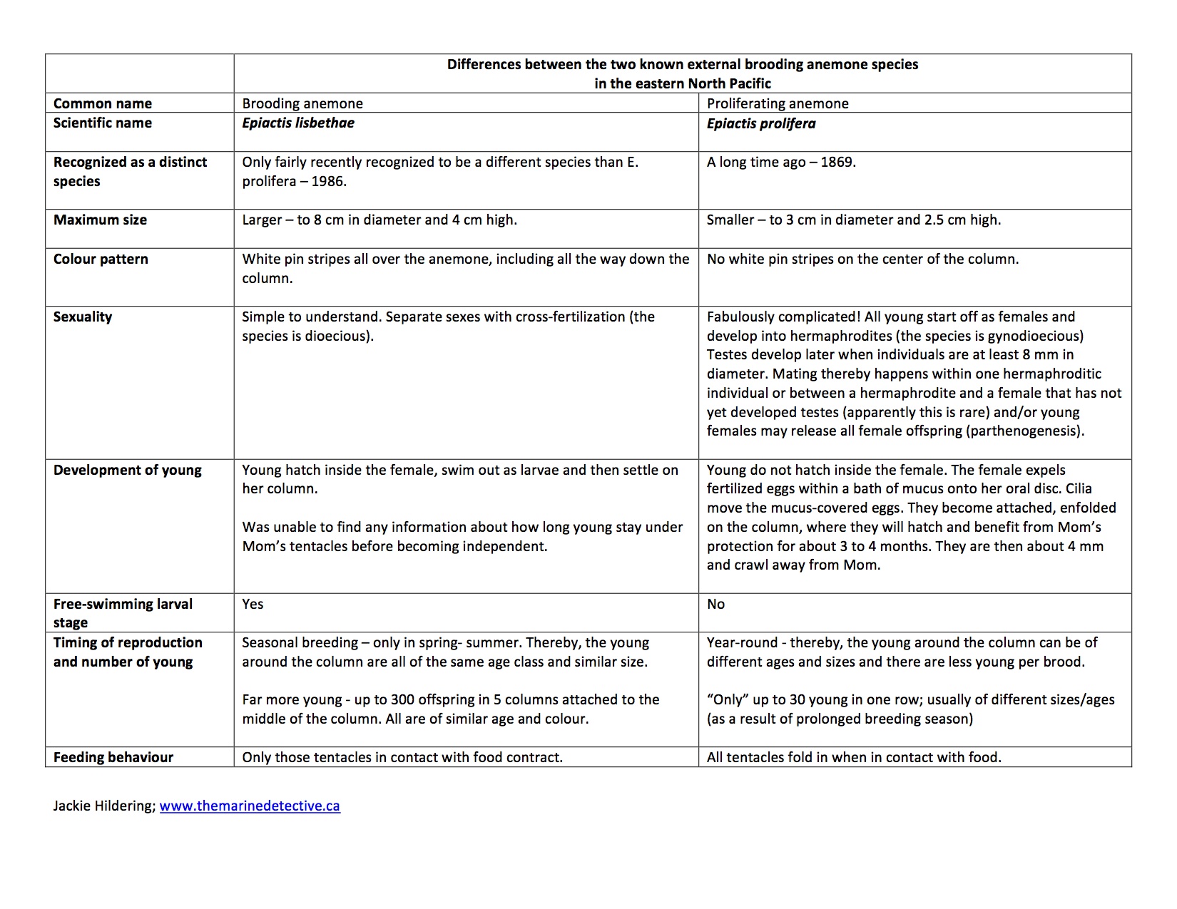
I have strived to clarify the many differences between these two externally brooding anemone species in the table below but to summarize: the Proliferating Anemone is smaller and does not have striping all the way down the column; adults are hermaphrodites; breeding happens year round; there are far fewer young clustered under mom’s tentacles; and they start off there as fertilized eggs, not as free-swimming larva.
The main similarity between these two species is and yes, I am going to use a tongue twister here since I believe it is inevitable when discussing anemones: with anemone mothers like these, baby anemones are protected from their anemone enemies!
Now off you go, share some ocean love with a Super Mom!
There are so many human females out there worthy of awe. Where, were we to consider how many young they have shielded and helped to independence, the number might well be 300 or more!
Click to enlarge. Table summarizing the differences between Brooding and Proliferating Anemones.
Next 3 photos show Proliferating Anemone babies under their mother’s tentacles, some shuffling off after 3 to 4 months there. Epiactis prolifera – often confused with the Brooding Anemone.
Updated with photos: 2025-12-18 An egg hunt mystery FINALLY came to an end for me, coincidentally, just before Easter when many of you were involved in egg hunts too.
I dare say however that my hunt involved vastly more beautiful eggs; that the hunt was much more challenging and – ultimately, much more rewarding!
One of my very, very favourite things to do, satisfying my “The Marine Detective” nature, is to solve the ultimate “whodunit” and match sea slug species with their egg masses / ribbons.
Every sea slug species’ egg mass is distinct, comprising a fascinating diversity of intoxicatingly beautiful shapes and patterns.
It delights me (for reasons I can’t fully explain) that for many sea slugs in the northeast Pacific Ocean, I am able to see an egg mass and immediately know which species laid it.
There are big clues because sea slugs most often lay eggs on their food. So if I know their prey preference I can narrow down which species laid the eggs.
Easiest of course is to have have the good fortune to find a sea slug in the act of laying their eggs.
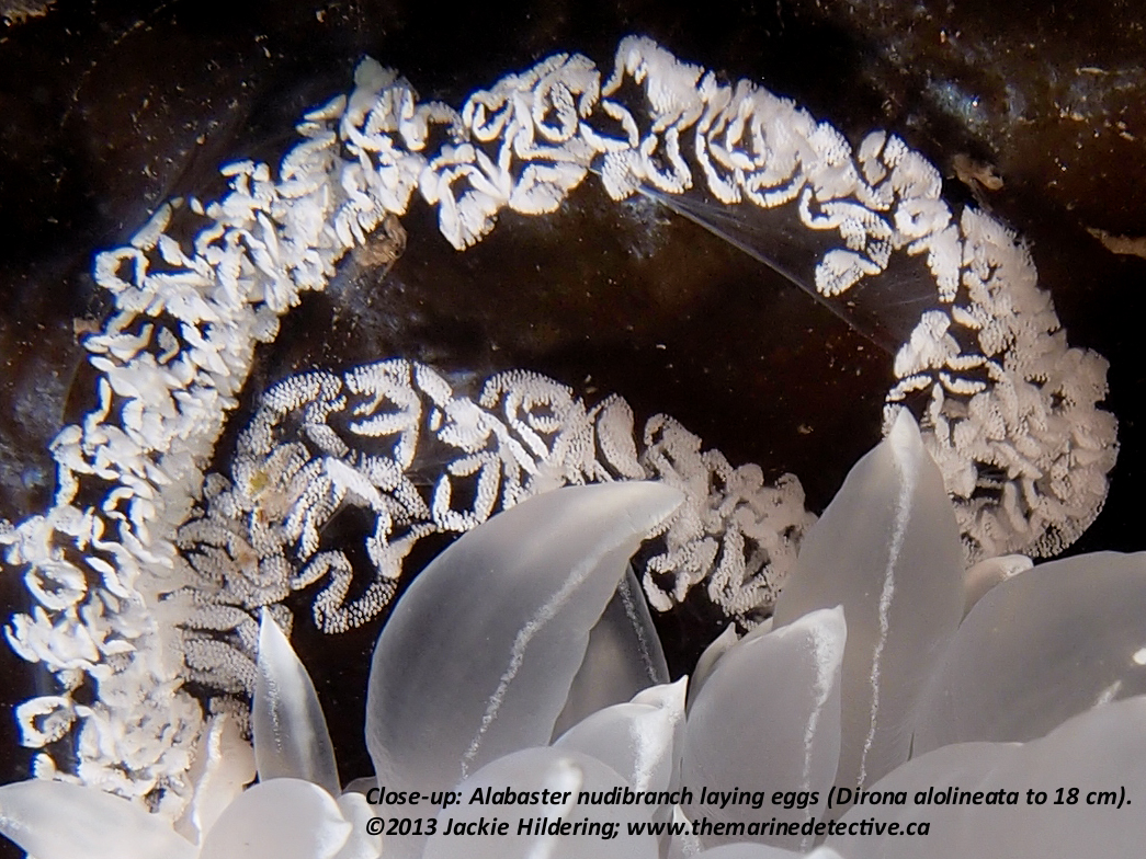
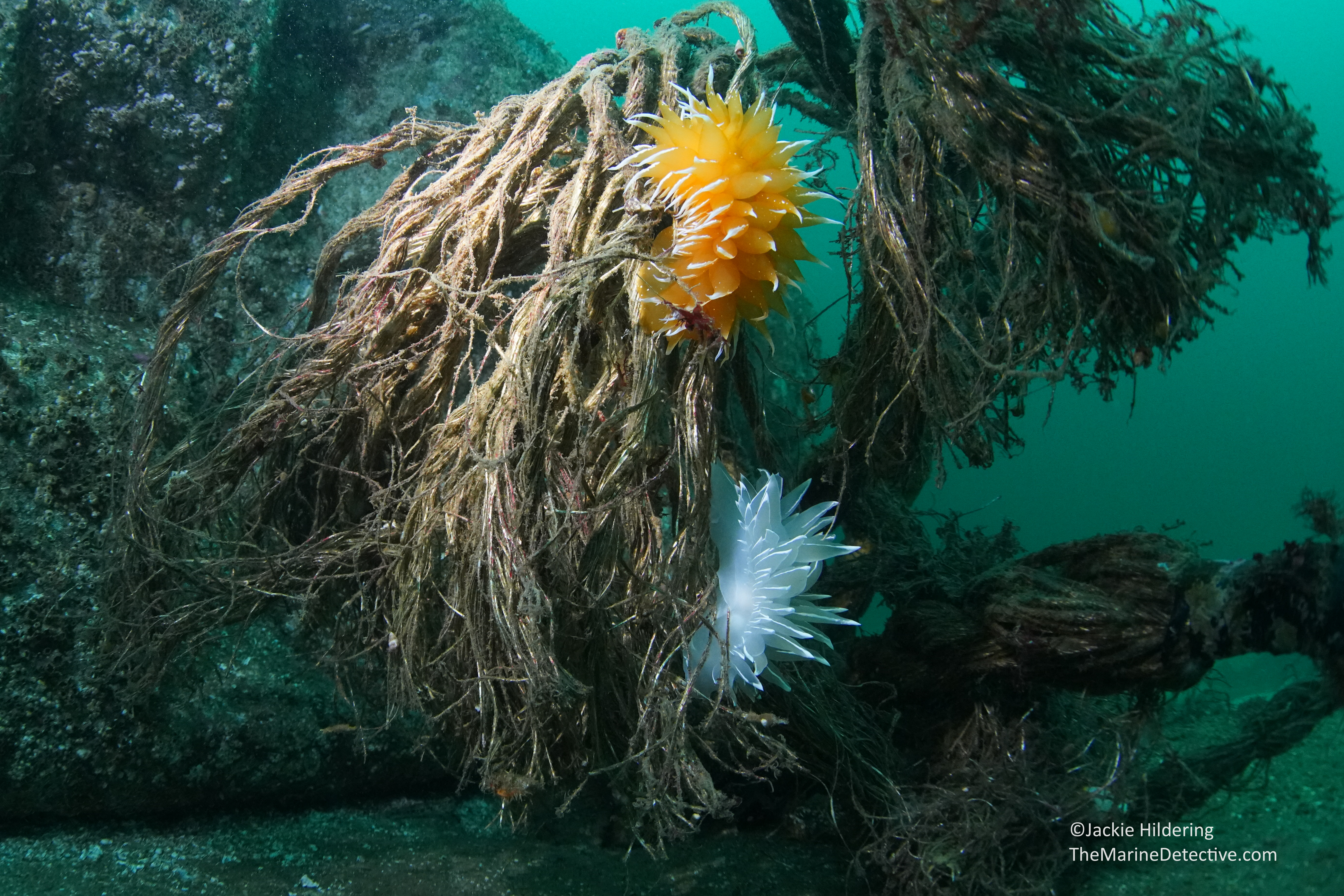
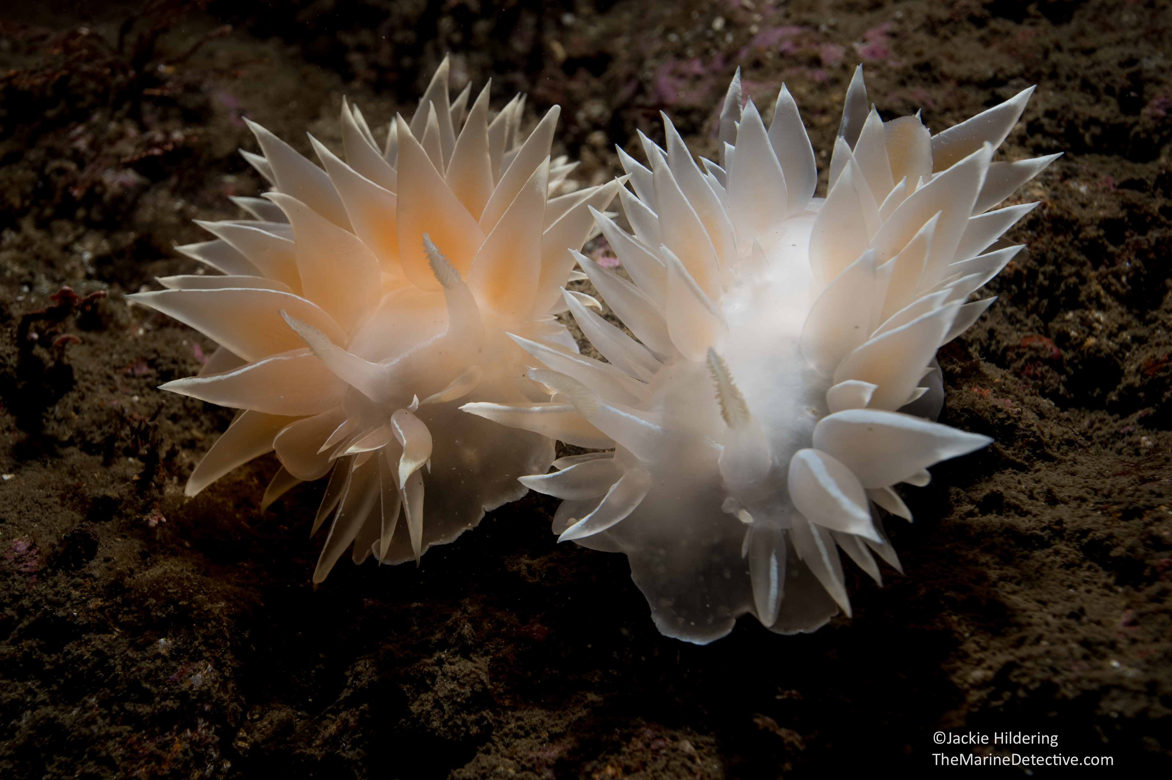
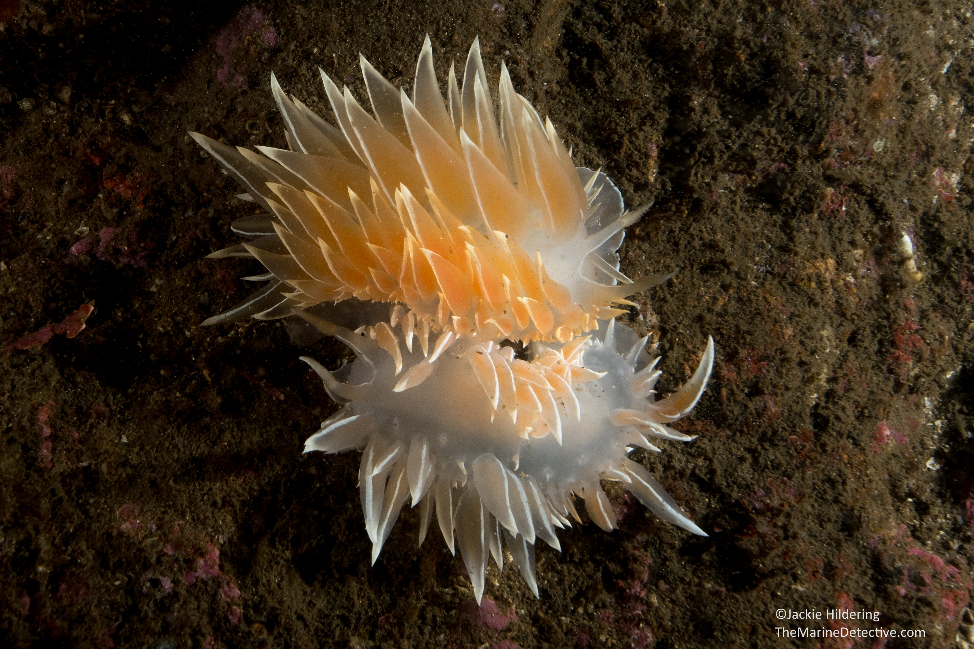
But for YEARS, I have been unable to differentiate the egg masses of two of the most beautiful sea slug species in these waters – the Gold Dirona (Dirona pellucida to 12 cm) and the Alabaster Nudibranch (Dirona albolineata to 18 cm and also referenced as the “White-Lined Dirona” or “Frosted Nudibranch”).
You’ll note that they are very closely related (same genus) and it is thereby not surprising that their egg masses would look very similar. Both also often lay their eggs on the same species of Agarum kelp. In all these years, while I have often found both species mating, I have never found either species laying their eggs.
But then, this week . . . just when I was noting the abundance of both species, how many egg masses there were and wishing, WISHING, I could find just one of them laying eggs – my dear dive buddy Jacqui Engel waved me over and pointed out a Gold Dirona laying eggs.
I was so jubilant, I screamed underwater. Yes, I am The Marine Detective for a reason, such things really do delight me to this degree.
Finally! Mystery solved, I would be able to differentiate the egg masses of the two species.
But then, Nature was even kinder to me.
On the very same day on the very same dive, after so many years, I also stumbled across an Alabaster Nudibranch laying eggs!
Disbelief! Joy! Manic photo-taking!
I think you may marvel at how very similar the masses are but the difference, at least to me is clear.
The “pieces” of the Gold Dirona’s egg mass are more compact and more like rice kernels.
The segments of the Alabaster Nudibranch’s egg masses are more scallop-edged and diffuse.
Please know that these differences would not be as clear if the eggs were older.
It’s estimated that there are ~350,000 eggs in one Alabaster Nudibranch egg mass. Source: WallaWallaEd. I do not know if this is the number of egg capsules (the dots you see), or if it includes the number of eggs in each egg capsule.
Sea slugs are reciprocal hermaphrodites which means that both become inseminated and lay eggs. One individual lays more than one egg mass as well. So many eggs are needed to ensure species survival when your young hatch out to become part of the planktonic soup of the Ocean.
If you have read to this point – thank you!
Likely we are kindred in our love of marine biodiversity and the beauty that is sea slugs.
For as much as I love chocolate Easter eggs, I would forego them for the rest of my existence if it would allow my appetite for marine mysteries to be further satisfied!
I’m excited to share video with you of Gumboot Chitons spawning. These marine neighbours most often seem quite inanimate – having a life where they keep their undersides protected by sucking down hard on rocks but, when it’s time to mate . . . . .
First just a little background: “Chitons” are marine molluscs (soft bodied animals) that, rather than having one of more shells to protect themselves, they have 8 armoured plates surrounded by a thick band of muscle. This allows them to suction onto surfaces very effectively since the 8-plates give such flexibility that they can even get a good grip on surfaces that are not flat.
There are many members of the the chiton class but the Gumboot Chiton (Cryptochiton steller; aka Giant Pacific Chiton) is very unique in its appearance.
It’s another “the biggest of its kind in the world” that inhabits the cold, rich waters of the northeast Pacific. It can be 35 cm long and about 2 kg. They are very slow growing and very long lived! This source reports that chitons that are 15 cm long are likely 20 years old and that they may live to be more than 25. That’s one old gumboot!
The Gumboot Chiton is also the only chiton species that has flesh completely covering the 8 plates. The texture and colour of this “girdle” offers them great camouflage and is where the “gumboot” descriptor comes from. The genus name “Cryptochiton” relates to this camouflage and that the 8 plates are hidden under the girdle. These plates are very uniquely shaped, and well-described with the name “butterfly shells”.
Apparently some First Nations did sometimes chew on this species but I am SURE that this is not the cultural origin of some people referring to this species as “wandering meatloaf”!
I don’t know where I picked this up, but I believe that one of the First Nations’ names for gumboot chitons translates (very) loosely, into “stuck on rock with face forever”. This would be an incredibly good descriptor since most chitons stay “face” down, grazing on algae by scraping with the sharp teeth-like structures of their radula. Thereby, they don’t expose their soft bodies and reduce the chance of predation.
I once found a Gumboot Chiton that had been dislodged by a predator at low tide. It is then that I learned that they have the ability to curl up on themselves like a pill bug!
But outside of a rare experience like this, you don’t often get a chance to see how very alive and animal-like they are.
Unless . . . they are spawning.
Then, up came the bodies of the Gumboot Chitons, into a very unique funnel-like shape. The “gonadal pores” are near the bottom end of the animals, but by positioning themselves in this shape, they channel the sex cells upward.
When spawning, you can clearly see which Gumboot Chitons are male and which are female!
It was just remarkable to see this, feeling truly as if some secret world was being revealed, and the coordinated timing of the spawning was astonishing.
Of course when you are a broadcast spawner, you need to release copious amounts of sex cells and need to do so at the same time or there will be even less chance that egg meets sperm. You can imagine how many eggs need to be fertilized if any of your zooplankton offspring are going to survive since so many animals feed on plankton.
To my knowledge, science has not concluded exactly what the cues are for “Hey fellow Gumbooot Chitons, it’s spawn time NOW!” It has to be temperature, light, tide and/or amount of food that determines the time is right.
Hum . . . seems to me that those cues may be significant between individuals of our species too!
Additional images:
Chiton plates on the right are those of the Gumboot Chiton.
I had a wonderful opportunity to photograph and film a lion’s mane jellyfish (Cyanea ferruginea) today.
The 1.5 minute annotated video clip below will give context to my “Sherlock – You Are Wrong” statement. Enjoy!
Click here to see a short clip of the other big jelly species that can be found in our waters – the egg yolk jelly (Phacellophora camtschatica) at up to “only” 60 cm across.
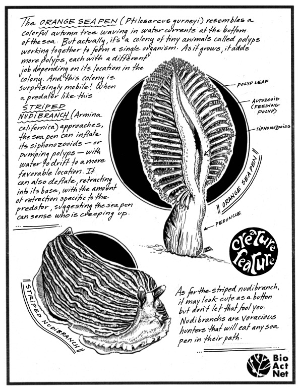
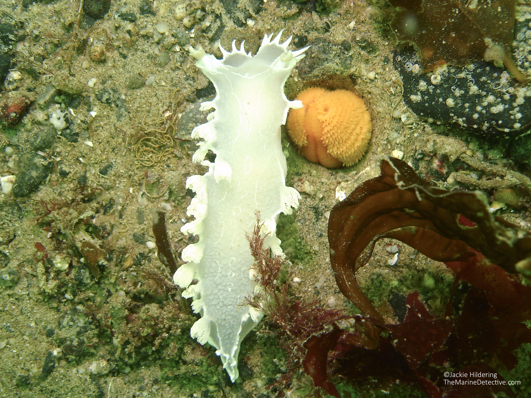
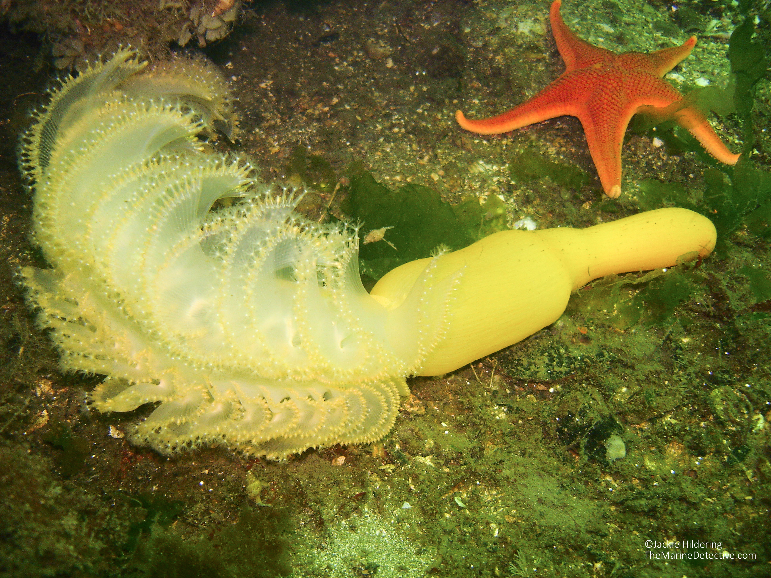
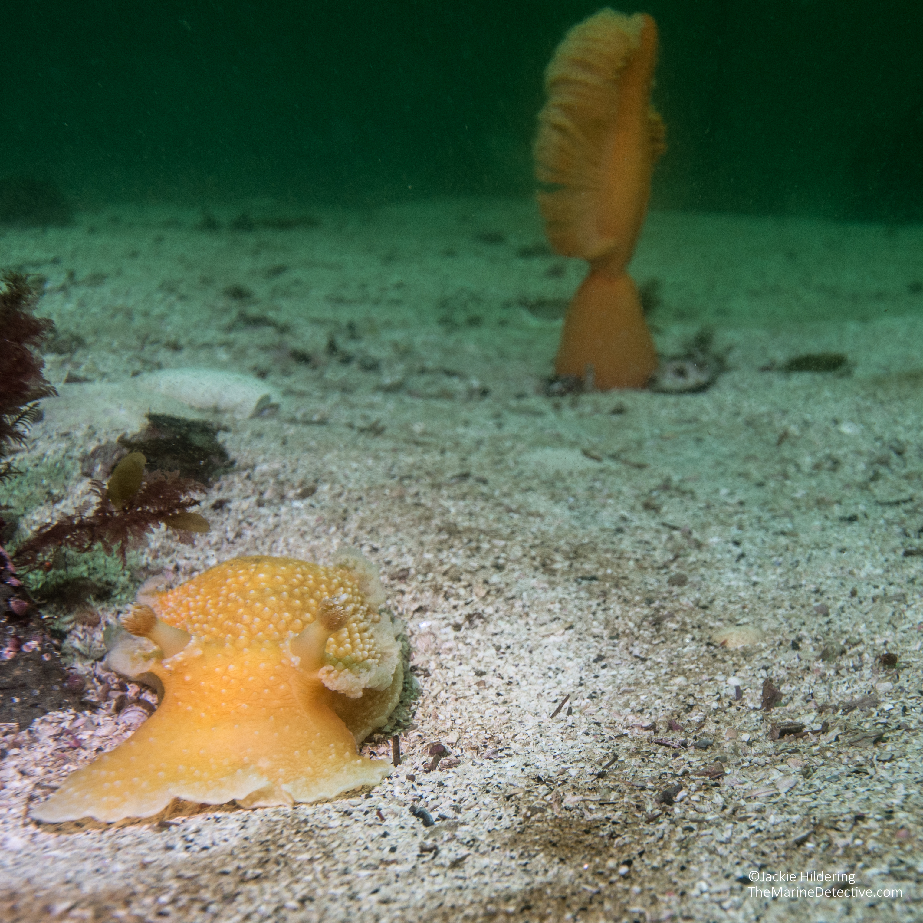
[Last updated November 15, 2023] This blog is about Sea Whips and Orange Sea Pens, the predators that stalk them, and how they can defend themselves.
These are the most surreal-looking organisms. Both species are octocorals – colonies of 8-tentacled polyp-like animals. The polyps filter feed on plankton.
Sea Whips can reach a height of 2.5 meters (Balticina willemoesi).Close-up on the feeding polyps of a Sea Whip. Orange Sea Pens can be up to 48 cm tall (Ptilosarcus gurney).Close-up on the feeding polyps of an Orange Sea Pen.
Information on Orange Sea Pens from the Monterey Bay Aquarium “A graceful creature of the seafloor, this sea pen resembles a plump, old-fashioned quill pen. Its colors range from dark orange to yellow to white. Each sea pen is a colony of polyps (small anemonelike individuals) working together for the survival of the whole. The primary polyp loses its tentacles and becomes the stalk of the sea pen, with a bulb at its base—the bulb anchors the sea pen in the muddy or sandy bottom. The various secondary polyps form the sea pen’s “branches” and have specialized functions. Some polyps feed by using nematocysts to catch plankton; some polyps reproduce; and some force water in and out of canals that ventilate the colony.”
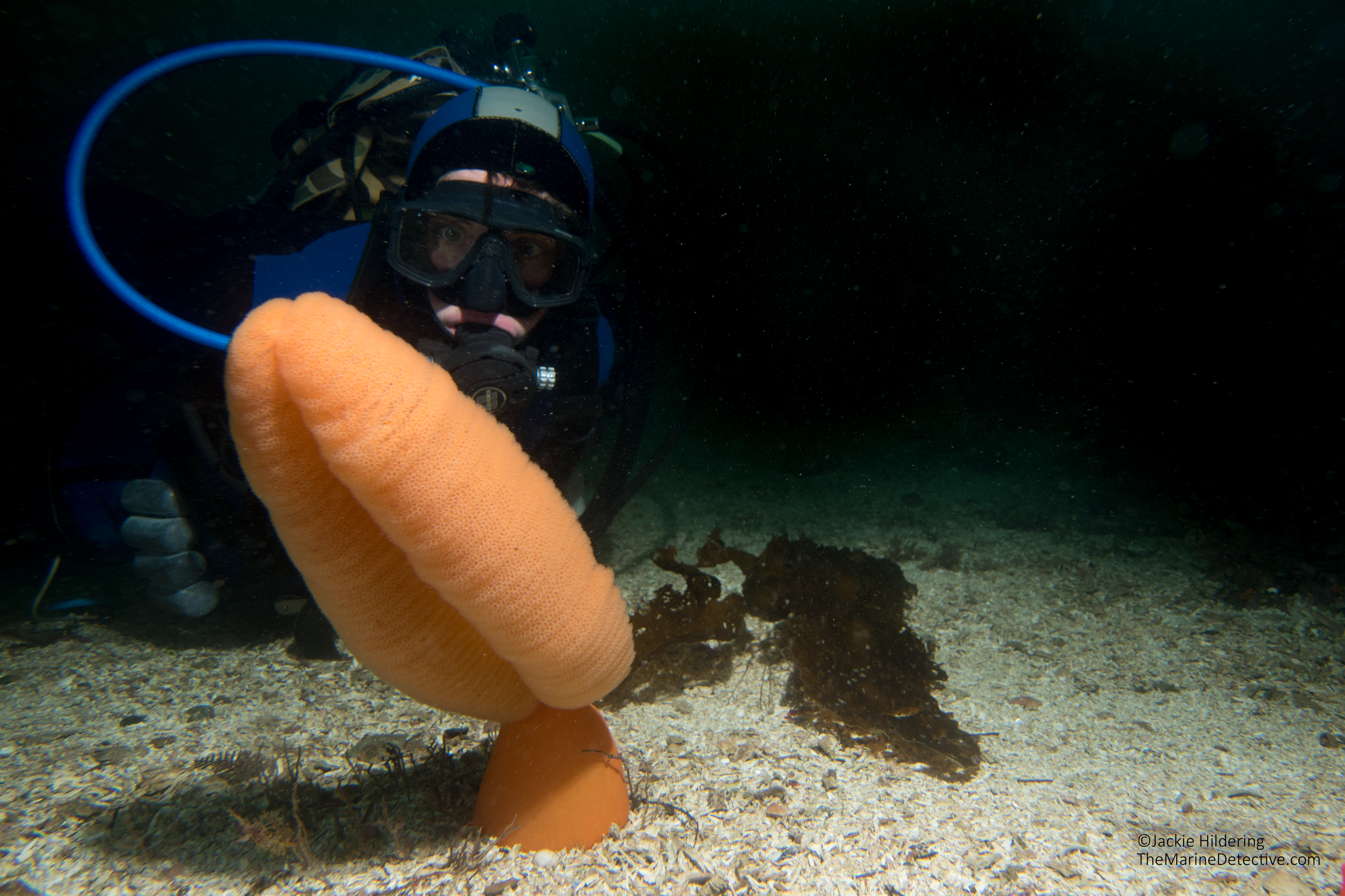
Dive buddy Natasha Dickinson with Orange Sea Pen
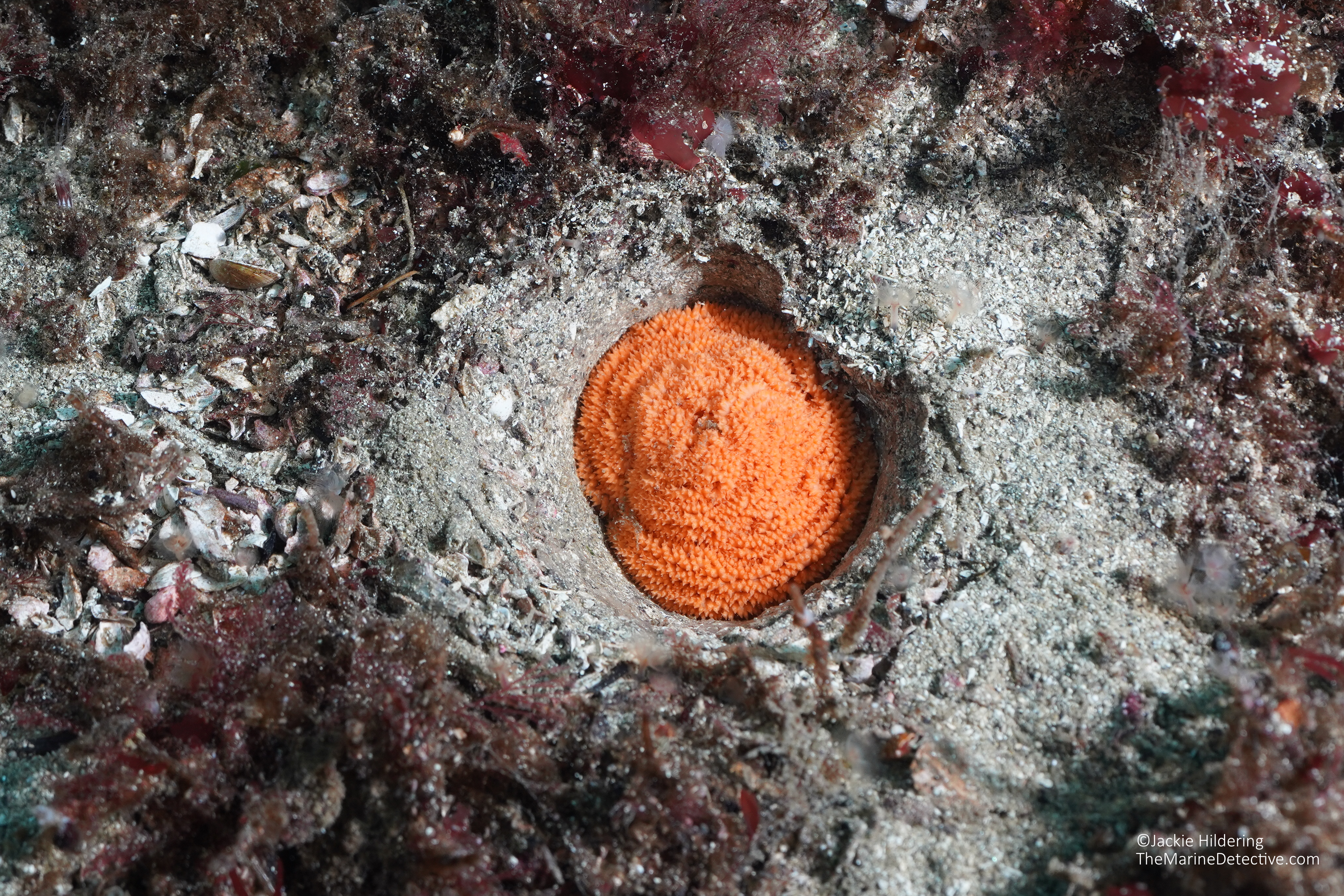
Remains of an Orange Sea Pen.
Orange Sea Pen Defences
When confronted by sea star and nudibranch predators, Orange Sea Pens can:
1) Deflate, “shrinking” down and into the sand. 2) Inflate, to drift away. 3) Create bioluminescence – making a greenish-blue light that is assumed to somehow deter predators. 4) Produce a toxin but this is poorly understood.
And you thought humans were special!
Deflated and retracted Orange Sea Pen. This can happen within about a minute of first contact from the predator (Wyeth & Willows, 2006 ). From “A Snail’s Odyssey: “A sea pen withdrawn into the sediment does not necessarily mean that it has been attacked or otherwise stimulated. Studies in Puget Sound, Washington show that sea pens Ptilosarcus gurneyi may inflate and deflate several times a day, and at any given time as few as one-quarter of all individuals are up and feeding.”
“The orange sea pen is surprisingly mobile, inflating its siphonozooids with water and drifting like a leaf on the wind when it wants to relocate. It can also deflate, partially retracting into its fleshy base when predators come calling. The amount of retraction has been shown to be specific to the approaching predator, which suggests that the pen can actually sense who is creeping up on it . . . Young sea pens are especially vulnerable to predation. They are incredibly slow-growing, taking over a year to reach about an inch tall. Orange Sea Pens increase their chances of survival with sheer numbers — a single pen can produce about a million eggs during its 10-year lifetime.”
Orange Sea Pen having a bad day? There are 4 predators here and it looks like the Orange Sea Pen was trying to inflate and drift away! But, there was a LOT of current whereby it kept being pushed down. Predators here are a Vermillion Star, two Diamondback Nudibranchs and one Orange Peel Nudibranch.
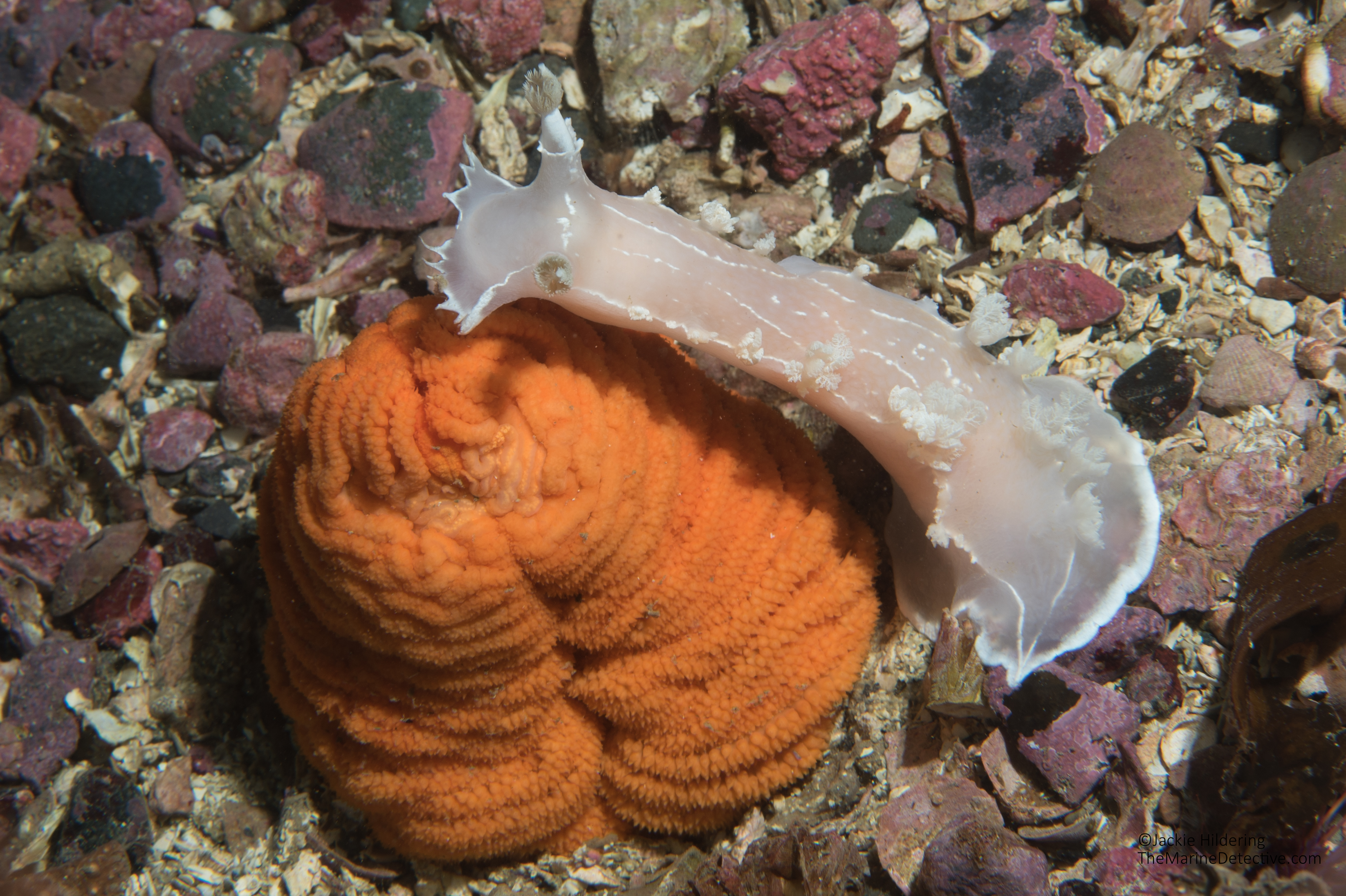
Orange Sea Pen being attacked by an Orange Peel Nudibranch.
Diamondback Nudibranch (predator) and a partially retracted Orange Sea Pen.
Striped Nudibranch feeding on the “pen” of an Orange Sea Pen. Giant Sea Cucumber on left.
By Romney McPhie. Click here for the PDF and more colouring sheets! Orange Sea Pen with inflated base. May have escaped predation this way – inflating and drifting away from the predator. Retracted Orange Sea Pen and Diamondback Nudibranch. Inflated Orange Sea Pen and Vermillion Star. Diamondback Nudibranch approaches its prey, the Orange Sea Pen. From the Washington State Department of Ecology: “The rows of feeding polyps on the feather-like structures “wave their 8 tentacles in the water to catch drifting plankton. These polyps [are] also responsible for producing eggs and sperm that get released into the water column. The siphonozooids, or pumping polyps, are found in the orange regions on the sides of the rachis [central stalk]. Their function is to take in or expel water, allowing the colony to inflate or deflate.”Diamondback Nudibranch crawling away from a retracted Orange Sea Pen. Orange Peel Nudibranch with Orange Sea Pen in the background.
The 2.5-minute clip below is of Sea Whips and Orange Sea Pens and the predators that stalk them.
Video: 1-minute of an Orange Sea Pen and Graceful Decorator Crab in the current in front of Port McNeill, BC.
The following BBC video below is from southwest Tasmania in Australia. This is not the sea pen species found in British Columbia. However, I have included it as it shows, with time-lapse photography, how sea pens can deflate and retract in response to predation attempts by nudibranchs.
Diamondback Nudibranch approaching and a White Sea Pen (Virgularia sp to 30 cm tall).
Note that I found very little information about the anti-predator responses of Sea Whips. From Malecha and Stone, 2009:
“For those colonies lying on the seafloor, most of the peduncles and the tissues of the rachis below the polyps (approximately 15 cm) were generally not consumed by Tritonia diomedea. Additionally, predation by nudibranchs on erect Halipteris willemoesi [now Balticina willemoesi] in the abraded and control groups did not occur. The disinclination of T. diomedea to prey on the lower portion of sea whips lying on the seafloor and their inability to prey on erect colonies perhaps suggests that H. willemoesi [Balticina willemoesi] may have structural and/or chemical defenses on the tissue above their peduncles that deter epibenthic predators. Typically, chemical defenses are concentrated at the distal portions of colonies where polyp density is greatest, whereas structural defenses are often concentrated near the base of colonies (Harvell & Fenical 1989, Wylie & Paul 1989). The distribution of defenses is perhaps an adaptation to various types of predators and their mode of attack. Therefore, if sea whips have evolved defensive structures located at the base of the colony that are specific to epibenthic, non-swimming predators such as nudibranchs and sea stars, the defenses certainly do not provide protection when sea whips are not erect. Disturbed animals, especially those lying on the seafloor, may be more vulnerable to predation from a wider array of predators since the defenses at their polyps may not be adaptively effective against non-swimming predators. Further study could examine the possible chemical and/or structural defenses of sea whips that are common among octocorals.”
It was 7.2° C (45° F) in the ocean yesterday. Even in summer, I’ve only experienced a high of about 10° C.
Typical for Northern Vancouver Island at this time of year, it was also windy enough for us to abort going out for a boat dive.
Windy, chilly, drizzly, grey . . . what’s a cold-water scuba diver to do?
Get in the cold, dark green water however you can because you KNOW what kind of beauty and wonder are always to be found below the surface, even where you moor your boat!
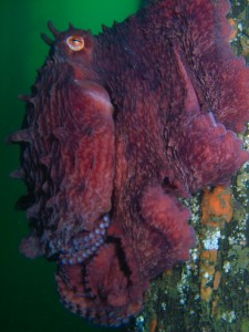
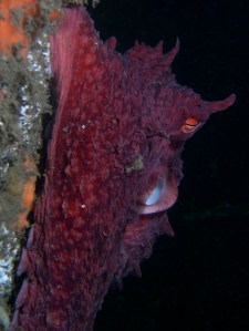
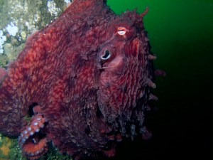
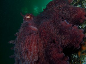
And indeed, under the dock, at only 6 m (20′) we found a Giant Pacific Octopus (Enteroctopus dofleini), curled up on a piling, incredibly tolerant of this ecstatic marine educator. Octopuses are SUCH intelligent animals. I felt as much like I was being scrutinized as he/she must have felt as I observed and photographed this awe-inspiring creature.
This individual was “only” about average size (23 to 42 kg). They can weigh more than 73 kg! I promise many more details on this species in future blog items.
There was so much other beauty under the dock but, for this blog posting, I will leave it at sharing the wonder of this Octo-brr octopus.
Bring on Novem-brrr to Fe-brrr-ary! The cold-water diving is so worth it!
To see these (and additional) images from this octopus photo-shoot at full size, click here.
I would greatly appreciate it if you would let me know, via blog comments, which image (#1 to 6) you believe is the best. This will determine which image ends up in next year’s WILD Northern Vancouver Island Calendar.

















 Sources: